 Explore
 Validate
 Learn
Learn Western blot
Antibody data
- Antibody Data
- Antigen structure
- References [1]
- Comments [0]
- Validations
- Western blot [3]
- Immunohistochemistry [2]
- Other assay [1]
Submit
Validation data
Reference
Comment
Report error
- Product number
- PA5-21394 - Provider product page

- Provider
- Invitrogen Antibodies
- Product name
- UQCRC1 Polyclonal Antibody
- Antibody type
- Polyclonal
- Antigen
- Recombinant protein fragment
- Description
- Recommended positive controls: 293T, Mouse brain.
- Concentration
- 1 mg/mL
Submitted references Initiation of electron transport chain activity in the embryonic heart coincides with the activation of mitochondrial complex 1 and the formation of supercomplexes.
Beutner G, Eliseev RA, Porter GA Jr
PloS one 2014;9(11):e113330
PloS one 2014;9(11):e113330
No comments: Submit comment
Supportive validation
- Submitted by
- Invitrogen Antibodies (provider)
- Main image
- Experimental details
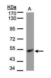
- Western blot analysis of ubiquinol-cytochrome c reductase core protein I precursor using 30 µg of HepG2 lysate. Samples were loaded onto a 7.5% SDS-PAGE gel and probed with a ubiquinol-cytochrome c reductase core protein I precursor polyclonal antibody (Product # PA5-21394) at a dilution of 1:1000.
- Submitted by
- Invitrogen Antibodies (provider)
- Main image
- Experimental details
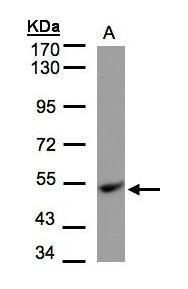
- Western Blot using UQCRC1 Polyclonal Antibody (Product # PA5-21394). Sample (50 µg of whole cell lysate). Lane A: Mouse brain. 10% SDS PAGE. UQCRC1 Polyclonal Antibody (Product # PA5-21394) diluted at 1:1,000.

- Submitted by
- Invitrogen Antibodies (provider)
- Main image
- Experimental details
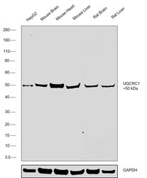
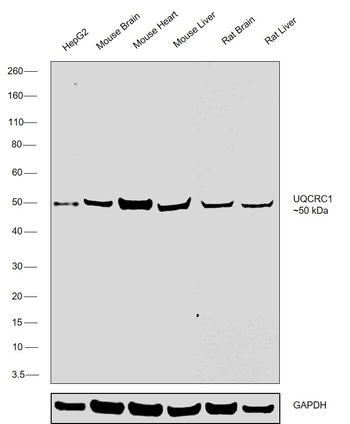
- Western blot was performed using UQCRC1 Polyclonal Antibody (Product # PA5-21394) and a ~50 kDa band corresponding to UQCRC1 was observed across cell line and tissues tested. Whole cell extracts (30 µg lysate) of HepG2 (Lane 1),tissue extracts (30 µg lysate) of Mouse Brain (Lane 2), Mouse Heart (Lane 3), Mouse Liver (Lane 4), Rat Brain (Lane 5) and Rat Liver (Lane 6) were electrophoresed using NuPAGE® 4-12 % Bis-Tris gel (Product # NP0321BOX). Resolved proteins were then transferred onto a nitrocellulose membrane (Product # IB23001) by iBlot® 2 Dry Blotting System (Product # IB21001). The blot was probed with the primary antibody (1:2000 dilution) and detected by chemiluminescence Goat anti-Rabbit IgG (H+L) Superclonal™ Recombinant Secondary Antibody, HRP (Product # A27036, 1:4000 dilution) using the iBright FL 1000 (Product # A32752). Chemiluminescent detection was performed using Novex® ECL Chemiluminescent Substrate Reagent Kit (Product # WP20005).
Supportive validation
- Submitted by
- Invitrogen Antibodies (provider)
- Main image
- Experimental details
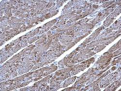
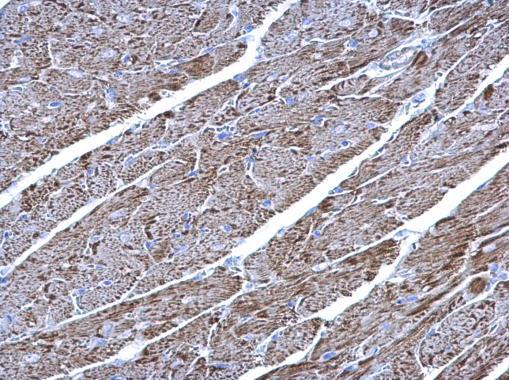
- UQCRC1 Polyclonal Antibody detects UQCRC1 protein at mitochondria on mouse heart by immunohistochemical analysis. Sample: Paraffin-embedded mouse heart. UQCRC1 Polyclonal Antibody (Product # PA5-21394) diluted at 1:500. Antigen Retrieval: EDTA based buffer, pH 8.0, 15 min.
- Submitted by
- Invitrogen Antibodies (provider)
- Main image

- Experimental details
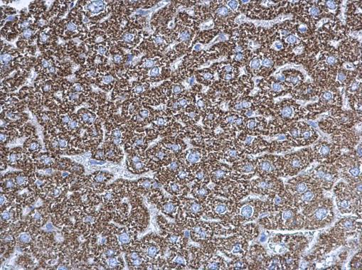
- UQCRC1 Polyclonal Antibody detects UQCRC1 protein at mitochondria on mouse liver by immunohistochemical analysis. Sample: Paraffin-embedded mouse liver. UQCRC1 Polyclonal Antibody (Product # PA5-21394) diluted at 1:500. Antigen Retrieval: EDTA based buffer, pH 8.0, 15 min.
Supportive validation
- Submitted by
- Invitrogen Antibodies (provider)
- Main image
- Experimental details
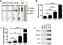
- Figure 8 The assembly of ETC complexes into supercomplexes begins around E11.5. A. One-dimensional high resolution clear native PAGE (hrCN-PAGE) shows the presence of monomeric Cx-1 and the appearance of Cx-1 containing supercomplexes during embryonic development and in adult mouse hearts. Whole tissue homogenates (10 ug for embryonic samples and 5 ug for adult sample) from hearts were solubilized with digitonin (4g digitonin/g protein) and separated by 5-15% hrCN-PAGE. The proteins were then transferred onto nitrocellulose membranes and Cx-1 was visualized first by the detection of NDUFAB1 (AB1, a rabbit polyclonal antibody). Then, the membrane was stripped and re-probed against NDUFB6 (B6, a mouse-monoclonal antibody). In addition, an in-gel assay of Cx-1 (IGA) demonstrates functional complex I monomers and supercomplexes are present in adult heart homogenates. B and C. Quantitative analysis of the presence of monomeric Cx-1 (B) and supercomplexes containing Cx-1 (C). NS-not significant, *p
