Explore
 Validate
 Learn
Learn Western blot
 Immunocytochemistry
Antibody data
- Antibody Data
- Antigen structure
- References [2]
- Comments [0]
- Validations
- Western blot [4]
- Immunocytochemistry [1]
- Immunohistochemistry [2]
Submit
Validation data
Reference
Comment
Report error
- Product number
- GTX628886 - Provider product page

- Provider
- GeneTex
- Product name
- COX4 antibody [GT6310]
- Antibody type
- Monoclonal
- Reactivity
- Human, Rat
- Host
- Mouse
Submitted references Lead (Pb) induced ATM-dependent mitophagy via PINK1/Parkin pathway.
Ca2+ influx-mediated dilation of the endoplasmic reticulum and c-FLIPL downregulation trigger CDDO-Me-induced apoptosis in breast cancer cells.
Gu X, Qi Y, Feng Z, Ma L, Gao K, Zhang Y
Toxicology letters 2018 Jul;291:92-100
Toxicology letters 2018 Jul;291:92-100
Ca2+ influx-mediated dilation of the endoplasmic reticulum and c-FLIPL downregulation trigger CDDO-Me-induced apoptosis in breast cancer cells.
Jeong SA, Kim IY, Lee AR, Yoon MJ, Cho H, Lee JS, Choi KS
Oncotarget 2015 Aug 28;6(25):21173-92
Oncotarget 2015 Aug 28;6(25):21173-92
No comments: Submit comment
Enhanced validation
Supportive validation
- Submitted by
- GeneTex (provider)
- Enhanced method
- Genetic validation
- Main image
- Experimental details
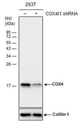
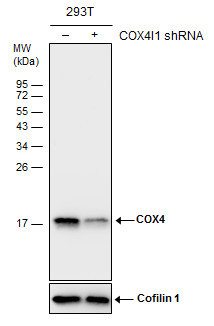
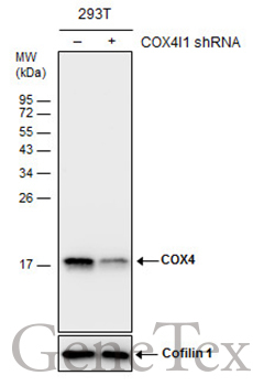
- Non-transfected (¡V) and transfected (+) 293T whole cell extracts (30 ?g) were separated by 15% SDS-PAGE, and the membrane was blotted with COX4 antibody [GT6310] (GTX628886) diluted at 1:1500.
Supportive validation
- Submitted by
- GeneTex (provider)
- Main image

- Experimental details
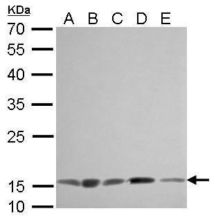
- COX4 antibody [GT6310] detects COX4I1 protein by Western blot analysis.A. 30 ?g 293T whole cell lysate/extractB. 30 ?g A431 whole cell lysate/extractC. 30 ?g HeLa whole cell lysate/extractD. 30 ?g HepG2 whole cell lysate/extractE. 30 ?g A375 whole cell lysate/extract12 % SDS-PAGECOX4 antibody [GT6310] (GTX628886) dilution: 1:1000
- Submitted by
- GeneTex (provider)
- Main image

- Experimental details
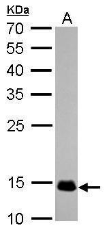
- COX4 antibody [GT6310] detects COX4I1 protein by Western blot analysis.A. 50 ?g rat muscle lysate/extract12 % SDS-PAGECOX4 antibody [GT6310] (GTX628886) dilution: 1:1000
- Submitted by
- GeneTex (provider)
- Main image

- Experimental details
- Non-transfected (¡V) and transfected (+) 293T whole cell extracts (30 ?g) were separated by 15% SDS-PAGE, and the membrane was blotted with COX4 antibody [GT6310] (GTX628886) diluted at 1:1500.
Supportive validation
- Submitted by
- GeneTex (provider)
- Main image

- Experimental details
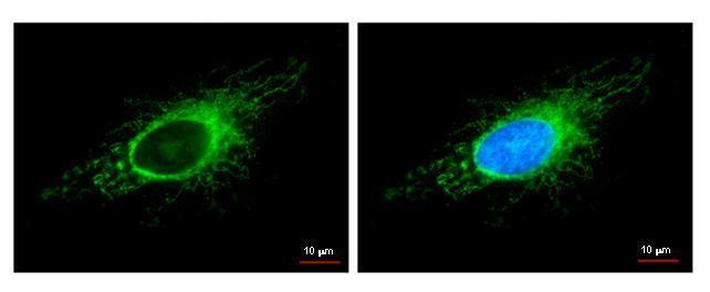
- COX4I1 antibody detects COX4I1 protein at mitochondria by immunofluorescent analysis. Sample: HeLa cells were fixed in -20¢J 100% methanol for 5 min.Green: COX4I1 protein stained by COX4I1 antibody (GTX628886) diluted at 1:500.Blue: Hoechst 33342 staining.
Supportive validation
- Submitted by
- GeneTex (provider)
- Main image

- Experimental details
- COX4 antibody [GT6310] detects COX4I1 protein at cytosol on U87 xenograft by immunohistochemical analysis. Sample: Paraffin-embedded U87 xenograft. COX4 antibody [GT6310] (GTX628886) dilution: 1:200.
- Submitted by
- GeneTex (provider)
- Main image

- Experimental details
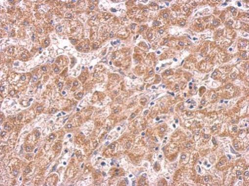
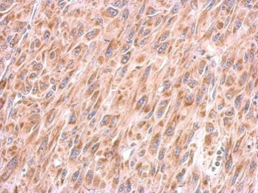
- COX4 antibody [GT6310] detects COX4I1 protein at cytosol on human hepatoma by immunohistochemical analysis. Sample: Paraffin-embedded hepatoma. COX4 antibody [GT6310] (GTX628886) dilution: 1:200.