 Explore
 Validate
 Learn
Learn Western blot
Antibody data
- Antibody Data
- Antigen structure
- References [0]
- Comments [0]
- Validations
- Western blot [4]
- Immunocytochemistry [2]
- Immunohistochemistry [2]
- Flow cytometry [1]
Submit
Validation data
Reference
Comment
Report error
- Product number
- 710333 - Provider product page

- Provider
- Invitrogen Antibodies
- Product name
- MMP16 Recombinant Polyclonal Antibody (13HCLC)
- Antibody type
- Polyclonal
- Antigen
- Recombinant full-length protein
- Description
- This antibody is predicted to react with mouse, rat, non-human primate and rabbit based on sequence homology.
- Antibody clone number
- 13HCLC
- Concentration
- 0.5 mg/mL
No comments: Submit comment
Supportive validation
- Submitted by
- Invitrogen Antibodies (provider)
- Main image
- Experimental details
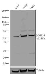
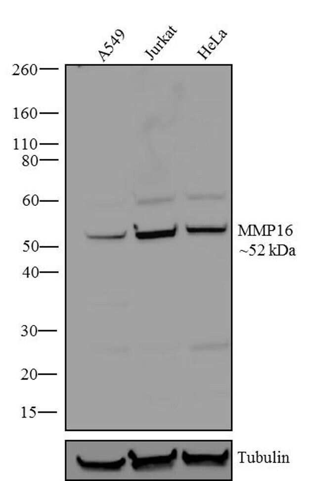
- Western blot analysis of MMP16 was performed by loading 20 µg of A549 (lane1), Jurkat (lane2) and HeLa (lane3) cell lysates using Novex®NuPAGE®4-12 % Bis-Tris gel (Product # NP0321BOX), XCell SureLock Electrophoresis System (Product # EI0002), Novex® Sharp Pre-Stained Protein Standard (Product # LC5800), and iBlot® Dry Blotting System (Product # IB21001). Proteins were transferred to a nitrocellulose membrane and blocked with 5 % skim milk for 1 hour at room temperature. MMP16 was detected at ~57 kDa using MMP16 Recombinant Rabbit Polyclonal Antibody (Product # 710333) at 1-3 µg/mL in 2.5 % skim milk at 4°C overnight on a rocking platform. Goat anti-Rabbit IgG-HRP Secondary Antibody (Product # G-21234) at 1:5000 dilution was used and chemiluminescent detection was performed using Pierce™ ECL Western blotting Substrate (Product # 32106).
- Submitted by
- Invitrogen Antibodies (provider)
- Main image

- Experimental details
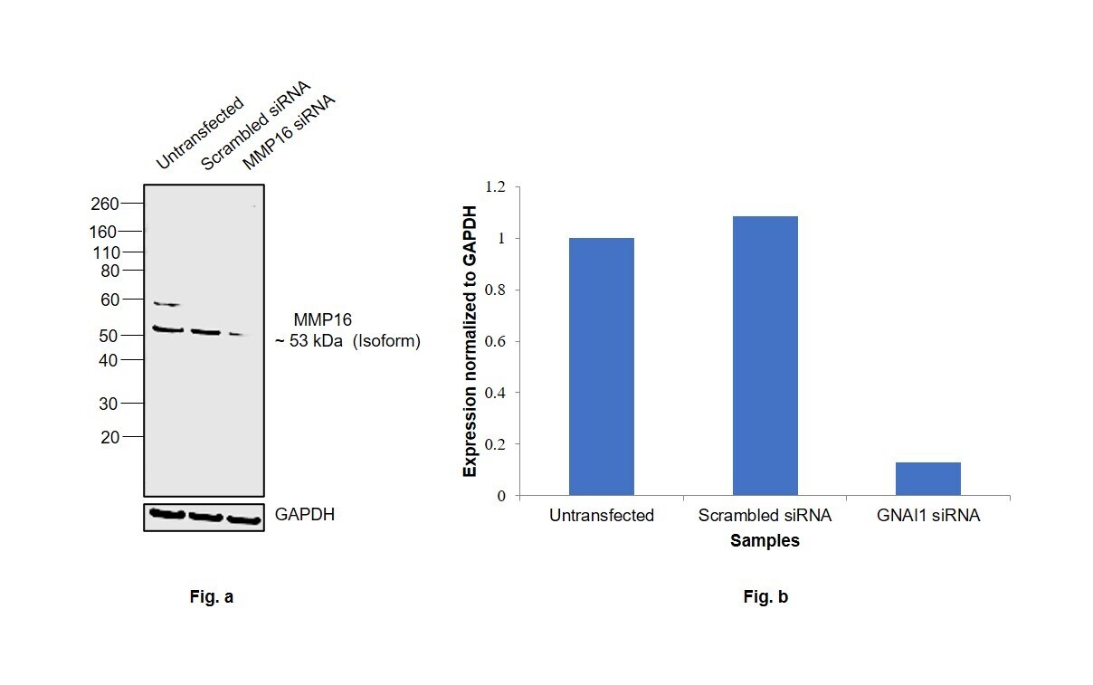
- Knockdown of Matrix metalloproteinase-16 was achieved by transfecting PC-3 with Matrix metalloproteinase-16 specific siRNAs (Silencer® select Product # s8883, s8885). Western blot analysis (Fig. a) was performed using Membrane enriched extracts from the Matrix metalloproteinase-16 knockdown cells (lane 3), non-targeting scrambled siRNA transfected cells (lane 2) and untransfected cells (lane 1). The blot was probed with MMP16 Recombinant Polyclonal Antibody (13HCLC) (Product # 710333, 1 µg/mL) and Goat anti-Rabbit IgG (H+L) Superclonal™ Recombinant Secondary Antibody, HRP (Product # A27036, 1:20000 dilution). Densitometric analysis of this western blot is shown in histogram (Fig. b). Decrease in signal upon siRNA mediated knock down confirms that antibody is specific to Matrix metalloproteinase-16 isoform.
- Submitted by
- Invitrogen Antibodies (provider)
- Main image
- Experimental details
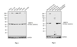
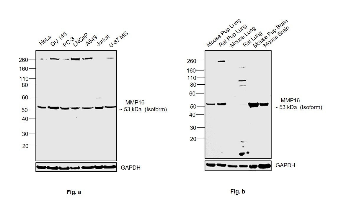
- Western blot was performed using Anti-MMP16 Recombinant Polyclonal Antibody (13HCLC) (Product # 710333) and a 53 kDa band corresponding to Matrix metalloproteinase-16 isoform was observed across cell lines and tissue extracts tested except Mouse Lung, Rat Lung and Mouse Bain which is reported to be low. Fig.a shows membrane enriched extracts (30 µg lysate) of HeLa (Lane 1), DU 145 (Lane 2), PC-3 (Lane 3), LNCaP (Lane 4), A549 (Lane 5), Jurkat (Lane 6), U-87 MG (Lane 7) and Fig.b shows tissue extracts (30 µg lysate) of Mouse Pup Lung (Lane 1), Rat Pup Lung (Lane 2), Mouse Lung (Lane 3), Rat Lung (Lane 4), Mouse Pup Brain (Lane 5) and Mouse Brain (Lane 6) were electrophoresed using NuPAGE™ 4-12% Bis-Tris Protein Gel (Product # NP0322BOX). Resolved proteins were then transferred onto a nitrocellulose membrane (Product # IB23001) by iBlot® 2 Dry Blotting System (Product # IB21001). The blot was probed with the primary antibody (1 µg/mL) and detected by chemiluminescence with Goat anti-Rabbit IgG (H+L) Superclonal™ Recombinant Secondary Antibody, HRP (Product # A27036,1:20000 dilution) using the iBright FL 1000 (Product # A32752). Chemiluminescent detection was performed using SuperSignal™ West Pico PLUS Chemiluminescent Substrate (Product # 34580).
- Submitted by
- Invitrogen Antibodies (provider)
- Main image

- Experimental details
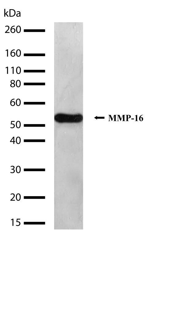
- Western blot analysis of MMP-16 in whole cells extracts from Jurkat cells using a MMP-16 Recombinant Rabbit Polyclonal Antibody (Product # 710333) at a dilution of 1 µg/mL. Samples were detected using chemiluminescence (ECL). Results show a band at ~52kDa.
Supportive validation
- Submitted by
- Invitrogen Antibodies (provider)
- Main image

- Experimental details
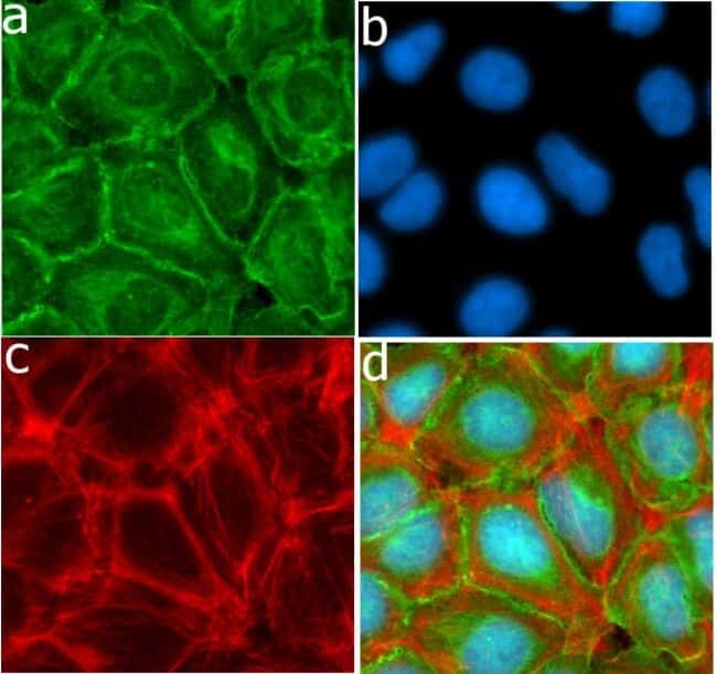
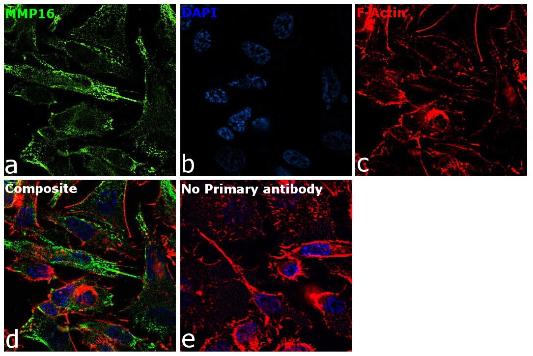
- Immunofluorescent analysis of MMP-16 in HeLa cells using a MMP-16 Recombinant Rabbit Polyclonal Antibody (Product # 710333) followed by detection using an Alexa Fluor 488-conjugated Goat anti-Rabbit secondary antibody (green) (Image A). Nuclei were stained using DAPI (Image B) and actin stained with Alexa Fluor 594 phalloidin (red) (image C). Image D is a composite image showing membrane localization of MMP-16.
- Submitted by
- Invitrogen Antibodies (provider)
- Main image
- Experimental details
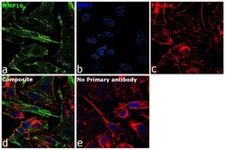
- Immunofluorescence analysis of Matrix metalloproteinase-16 was performed using 70% confluent log phase PC-3 cells. The cells were fixed with 4% paraformaldehyde for 10 minutes, permeabilized with 0.1% Triton™ X-100 for 15 minutes, and blocked with 2% BSA for 45 minutes at room temperature. The cells were labeled with MMP16 Recombinant Polyclonal Antibody (13HCLC) (Product # 710333) at 1 µg/mL in 0.1% BSA, incubated at 4 degree celsius overnight and then labeled with Donkey anti-Rabbit IgG (H+L) Highly Cross-Adsorbed Secondary Antibody, Alexa Fluor Plus 488 (Product # A32790), (1:2000), for 45 minutes at room temperature (Panel a: Green). Nuclei (Panel b:Blue) were stained with ProLong™ Diamond Antifade Mountant with DAPI (Product # P36962). F-actin (Panel c: Red) was stained with Rhodamine Phalloidin (Product # R415, 1:300). Panel d represents the merged image showing cytoplasmic localization. Panel e represents control cells with no primary antibody to assess background. The images were captured at 60X magnification.
Supportive validation
- Submitted by
- Invitrogen Antibodies (provider)
- Main image
- Experimental details
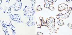
- Immunohistochemistry analysis of MMP-16 showing staining in the membrane of paraffin-embedded human placenta tissue (right) compared to a negative control without primary antibody (left). To expose target proteins, antigen retrieval was performed using 10 mM sodium citrate (pH 6.0), microwaved for 8-15 min. Following antigen retrieval, tissues were blocked in 3% H2O2-methanol for 15 min at room temperature, washed with ddH2O and PBS, and then probed with MMP-16 (13HCLC) Recombinant Rabbit Polyclonal Antibody (Product # 710333) diluted in 3% BSA-PBS at a dilution of 1:100 overnight at 4°C in a humidified chamber. Tissues were washed extensively in PBST and detection was performed using a HRP-conjugated secondary antibody followed by colorimetric detection using a DAB kit. Tissues were counterstained with hematoxylin and dehydrated with ethanol and xylene to prep for mounting.
- Submitted by
- Invitrogen Antibodies (provider)
- Main image

- Experimental details
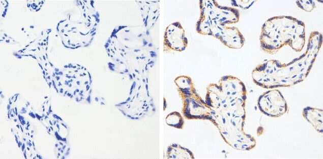
- Immunohistochemistry analysis of MMP-16 showing staining in the membrane of paraffin-embedded human placenta tissue (right) compared to a negative control without primary antibody (left). To expose target proteins, antigen retrieval was performed using 10 mM sodium citrate (pH 6.0), microwaved for 8-15 min. Following antigen retrieval, tissues were blocked in 3% H2O2-methanol for 15 min at room temperature, washed with ddH2O and PBS, and then probed with MMP-16 (13HCLC) Recombinant Rabbit Polyclonal Antibody (Product # 710333) diluted in 3% BSA-PBS at a dilution of 1:100 overnight at 4°C in a humidified chamber. Tissues were washed extensively in PBST and detection was performed using a HRP-conjugated secondary antibody followed by colorimetric detection using a DAB kit. Tissues were counterstained with hematoxylin and dehydrated with ethanol and xylene to prep for mounting.

Supportive validation
- Submitted by
- Invitrogen Antibodies (provider)
- Main image
- Experimental details
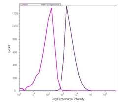
- Flow cytometry analysis of MMP-16 in HeLa cells using a MMP-16 Recombinant Rabbit Polyclonal Antibody (Product # 710333). Cells were fixed and permeabilized using FIX & PERM (Product # GAS-004) reagent, and detection was performed using an Alexa Fluor 488 Goat anti-Rabbit IgG (right peak) compared to a control without primary antibody (left peak).
