Explore
 Validate
 Learn
Learn Western blot
 Immunocytochemistry
Antibody data
- Antibody Data
- Antigen structure
- References [3]
- Comments [0]
- Validations
- Western blot [5]
- Immunoprecipitation [1]
Submit
Validation data
Reference
Comment
Report error
- Product number
- NB300-750 - Provider product page

- Provider
- Novus Biologicals
- Proper citation
- Novus Cat#NB300-750, RRID:AB_791502
- Product name
- Rabbit Polyclonal Pax6 Antibody
- Antibody type
- Polyclonal
- Description
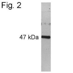
- Immunogen affinity purified. Detects a ~47 kDa protein representing PAX6 (Schwann Cell Marker) from rat whole eye extracts and ovine retinal extracts.
- Reactivity
- Human, Mouse, Rat, Sheep, Simian
- Host
- Rabbit
- Isotype
- IgG
- Vial size
- 100 ug
- Concentration
- 1 mg/ml
- Storage
- Store at -20C. Avoid freeze-thaw cycles.
Submitted references Evidence for generative homology of cerebellum and cerebellum-like structures in an elasmobranch fish based on Pax6, Cbln1 and Grid2 expression.
Renal progenitors derived from human iPSCs engraft and restore function in a mouse model of acute kidney injury.
Mapping differentiation kinetics in the mouse retina reveals an extensive period of cell cycle protein expression in post-mitotic newborn neurons.
Suriano CM, Bodznick D
The Journal of comparative neurology 2018 Oct 1;526(14):2187-2203
The Journal of comparative neurology 2018 Oct 1;526(14):2187-2203
Renal progenitors derived from human iPSCs engraft and restore function in a mouse model of acute kidney injury.
Imberti B, Tomasoni S, Ciampi O, Pezzotta A, Derosas M, Xinaris C, Rizzo P, Papadimou E, Novelli R, Benigni A, Remuzzi G, Morigi M
Scientific reports 2015 Mar 6;5:8826
Scientific reports 2015 Mar 6;5:8826
Mapping differentiation kinetics in the mouse retina reveals an extensive period of cell cycle protein expression in post-mitotic newborn neurons.
Pacal M, Bremner R
Developmental dynamics : an official publication of the American Association of Anatomists 2012 Oct;241(10):1525-44
Developmental dynamics : an official publication of the American Association of Anatomists 2012 Oct;241(10):1525-44
No comments: Submit comment
Supportive validation
- Submitted by
- Novus Biologicals (provider)
- Main image
- Experimental details
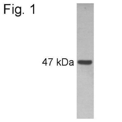
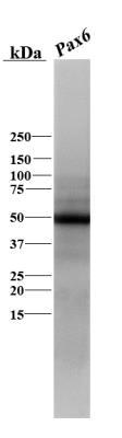
- Western Blot: Pax6 Antibody [NB300-750] - Analysis of rat whole eye extract
- Submitted by
- Novus Biologicals (provider)
- Main image

- Experimental details
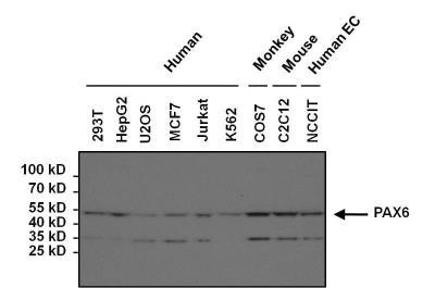
- Western Blot: Pax6 Antibody [NB300-750] - Analysis of various human, mouse and non-human primate whole cell lysates.
- Submitted by
- Novus Biologicals (provider)
- Main image
- Experimental details
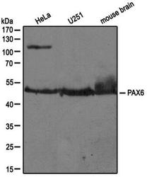
- Western Blot: Pax6 Antibody [NB300-750] - Analysis of extracts from HeLa, U251 and mouse brain using PAX6 antibody (NB300-750, 1:100).

- Submitted by
- Novus Biologicals (provider)
- Main image

- Experimental details
- Western Blot: Pax6 Antibody [NB300-750] - Analysis of Pax6 in skate (Leucoraja erinacea) brain lysate using anti-Pax6 antibody. Image from verified customer review.
- Submitted by
- Novus Biologicals (provider)
- Main image
- Experimental details
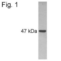
- Western Blot: Pax6 Antibody [NB300-750] - Analysis of sheep retinal extract

Supportive validation
- Submitted by
- Novus Biologicals (provider)
- Main image

- Experimental details
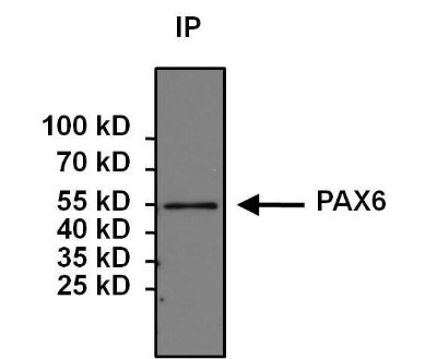
- Immunoprecipitation: Pax6 Antibody [NB300-750] - Analysis of PAX6 was performed on 293T cells. Antigen: antibody complexes were formed by incubating 500ug whole cell lysate with 5ug of PAX6 polyclonal antibody overnight on a rocking platform at 4C. The immune complexes were captured on 50ul Protein A/G Plus Agarose, washed extensively, and eluted with 5X Lane Marker Reducing Sample Buffer. Samples were resolved on a 4-20% Tris-HCl polyacrylamide gel, transferred to a PVDF membrane, and blocked with 5% BSA/TBS-0.1%Tween for at least 1 hour. The membrane was probed with a PAX6 monoclonal antibody at a dilution of 1:1000 overnight rotating at 4C, washed in TBST, and probed with Clean-Blot IP Detection Reagent at a dilution of 1:1000 for at least one hour.