 Explore
 Validate
 Learn
Learn Western blot
Antibody data
- Antibody Data
- Antigen structure
- References [4]
- Comments [0]
- Validations
- Western blot [3]
- Immunocytochemistry [2]
- Other assay [4]
Submit
Validation data
Reference
Comment
Report error
- Product number
- PA1-801 - Provider product page

- Provider
- Invitrogen Antibodies
- Product name
- PAX6 Polyclonal Antibody
- Antibody type
- Polyclonal
- Antigen
- Synthetic peptide
- Description
- PA1-801 detects PAX6 from human, mouse, rat, non-human primate and ovine samples. This antibody does not detect bovine PAX6 by Western blot.
- Concentration
- 1 mg/mL
Submitted references Core transcription regulatory circuitry orchestrates corneal epithelial homeostasis.
Effect of Ionizing Radiation from Computed Tomography on Differentiation of Human Embryonic Stem Cells into Neural Precursors.
Sonic hedgehog signalling regulates the self-renewal and proliferation of skin-derived precursor cells in mice.
Induced pluripotency enables differentiation of human nullipotent embryonal carcinoma cells N2102Ep.
Li M, Huang H, Li L, He C, Zhu L, Guo H, Wang L, Liu J, Wu S, Liu J, Xu T, Mao Z, Cao N, Zhang K, Lan F, Ding J, Yuan J, Liu Y, Ouyang H
Nature communications 2021 Jan 18;12(1):420
Nature communications 2021 Jan 18;12(1):420
Effect of Ionizing Radiation from Computed Tomography on Differentiation of Human Embryonic Stem Cells into Neural Precursors.
Hanu C, Loeliger BW, Panyutin IV, Maass-Moreno R, Wakim P, Pritchard WF, Neumann RD, Panyutin IG
International journal of molecular sciences 2019 Aug 10;20(16)
International journal of molecular sciences 2019 Aug 10;20(16)
Sonic hedgehog signalling regulates the self-renewal and proliferation of skin-derived precursor cells in mice.
Park S, Kim H, Kim K, Roh S
Cell proliferation 2018 Dec;51(6):e12500
Cell proliferation 2018 Dec;51(6):e12500
Induced pluripotency enables differentiation of human nullipotent embryonal carcinoma cells N2102Ep.
Sutiwisesak R, Kitiyanant N, Kotchabhakdi N, Felsenfeld G, Andrews PW, Wongtrakoongate P
Biochimica et biophysica acta 2014 Nov;1843(11):2611-9
Biochimica et biophysica acta 2014 Nov;1843(11):2611-9
No comments: Submit comment
Supportive validation
- Submitted by
- Invitrogen Antibodies (provider)
- Main image
- Experimental details
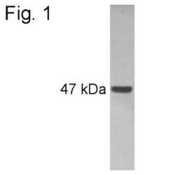
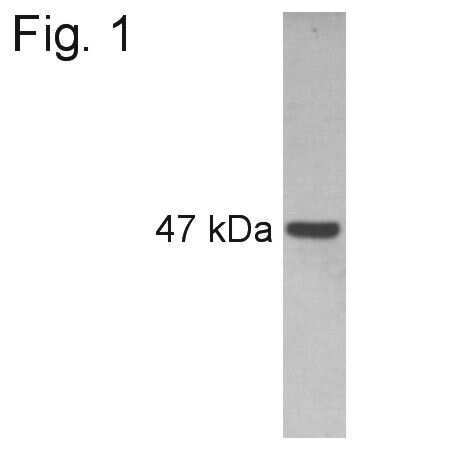
- Western blot analysis of PAX6 on rat whole eye extract using a PAX6 polyclonal antibody (Product # PA1-801).
- Submitted by
- Invitrogen Antibodies (provider)
- Main image

- Experimental details
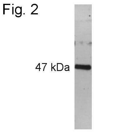
- Western blot analysis of PAX6 on ovine retinal extract using a PAX6 polyclonal antibody (Product # PA1-801).
- Submitted by
- Invitrogen Antibodies (provider)
- Main image
- Experimental details
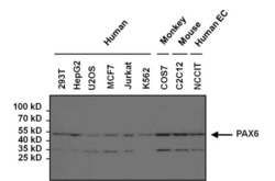
- Western blot analysis of PAX6 was performed by loading 50 µg of various human, mouse and non-human primate whole cell lysates per well onto a 4-20% Tris-HCl polyacrylamide gel. Proteins were transferred to a PVDF membrane and blocked with 5% BSA/TBST for at least 1 hour. The membrane was probed with a PAX6 polyclonal antibody (Product # PA1-801) at a dilution of 1:1000 overnight at 4°C on a rocking platform, washed in TBS-0.1%Tween 20, and probed with a goat anti-rabbit IgG-HRP secondary antibody (Product # 31460) at a dilution of 1:20,000 for at least one hour. Chemiluminescent detection was performed using SuperSignal West Dura (Product # 34075).

Supportive validation
- Submitted by
- Invitrogen Antibodies (provider)
- Main image
- Experimental details
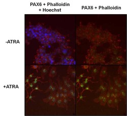
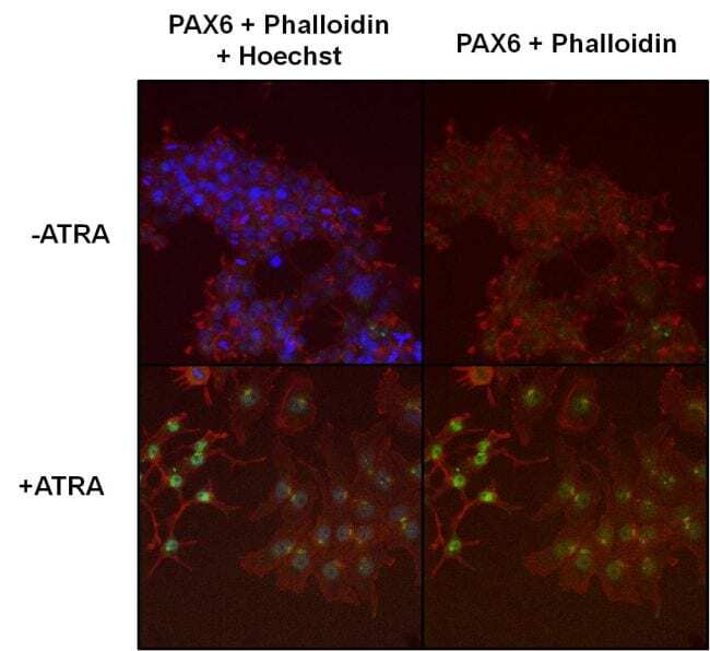
- Immunofluorescent analysis of PAX6 (green) in All-Trans-Retinoic Acid (ATRA) treated (10µM, 24 hours) NCCIT cells. Formalin fixed cells were permeabilized with 0.1% Triton X-100 in TBS for 10 minutes at room temperature and blocked with 1% Blocker BSA (Product # 37525) for 15 minutes at room temperature. Cells were probed with a PAX6 polyclonal antibody (Product # PA1-801), at a dilution of 1:50 for at least 1 hour at room temperature, washed with PBS, and incubated with DyLight 488 goat-anti-rabbit IgG secondary antibody (Product # 35552) at a dilution of 1:400 for 30 minutes at room temperature. F-Actin (red) was stained with DyLight 554 Phalloidin (Product # 21834), and nuclei (blue) were stained with Hoechst 33342 dye (Product # 62249). Images were taken on a Thermo Scientific ArrayScan at 20X magnification.

- Submitted by
- Invitrogen Antibodies (provider)
- Main image
- Experimental details
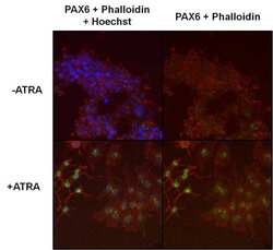
- Immunofluorescent analysis of PAX6 (green) in All-Trans-Retinoic Acid (ATRA) treated (10µM, 24 hours) NCCIT cells. Formalin fixed cells were permeabilized with 0.1% Triton X-100 in TBS for 10 minutes at room temperature and blocked with 1% Blocker BSA (Product # 37525) for 15 minutes at room temperature. Cells were probed with a PAX6 polyclonal antibody (Product # PA1-801), at a dilution of 1:50 for at least 1 hour at room temperature, washed with PBS, and incubated with DyLight 488 goat-anti-rabbit IgG secondary antibody (Product # 35552) at a dilution of 1:400 for 30 minutes at room temperature. F-Actin (red) was stained with DyLight 554 Phalloidin (Product # 21834), and nuclei (blue) were stained with Hoechst 33342 dye (Product # 62249). Images were taken on a Thermo Scientific ArrayScan at 20X magnification.
Supportive validation
- Submitted by
- Invitrogen Antibodies (provider)
- Main image
- Experimental details
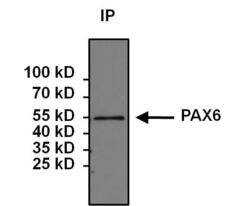
- Immunoprecipitation of PAX6 was performed on 293T cells. Antigen: antibody complexes were formed by incubating 500 µg whole cell lysate with 5 µg of PAX6 polyclonal antibody (Product # PA1-801) overnight on a rocking platform at 4°C. The immune complexes were captured on 50 µL Protein A/G Plus Agarose (Product # 20423), washed extensively, and eluted with 5X Lane Marker Reducing Sample Buffer (Product # 39000). Samples were resolved on a 4-20% Tris-HCl polyacrylamide gel, transferred to a PVDF membrane, and blocked with 5% BSA/TBS-0.1%Tween for at least 1 hour. The membrane was probed with a PAX6 monoclonal antibody (Product # PA1-801) at a dilution of 1:1000 overnight rotating at 4°C, washed in TBST, and probed with Clean-Blot IP Detection Reagent (Product # 21230) at a dilution of 1:1000 for at least one hour. Chemiluminescent detection was performed using SuperSignal West Dura (Product # 34075).

- Submitted by
- Invitrogen Antibodies (provider)
- Main image
- Experimental details
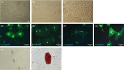
- Figure 1 Isolation and differentiation of murine skin-derived precursors ( mSKP s). (A) Single cells were separated from the back skin of murine foetuses. (B) Primary spheres were generated from isolated single cells. (C) Secondary spheres were larger and more condensed than primary spheres. (D-G) The mSKP s were differentiated into Schwann cells on PDL and laminin-coated dishes from d 14 to 21, using Schwann cell differentiation medium. The differentiated cells exhibited immunofluorescence (green) for (D) nerve growth factor receptor ( NGFR ), (E) myelin basic protein ( MBP ), (F) S100 calcium-binding protein (S100B) and (G) neural crest marker PAX 6. DAPI (blue) is shown in merged images (D-G). (H, I) The mSKP s were differentiated into adipogenic cells. The differentiated adipogenic cells from mSKP s were stained by Oil Red O. Scale bars: 100 mum

- Submitted by
- Invitrogen Antibodies (provider)
- Main image
- Experimental details
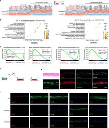
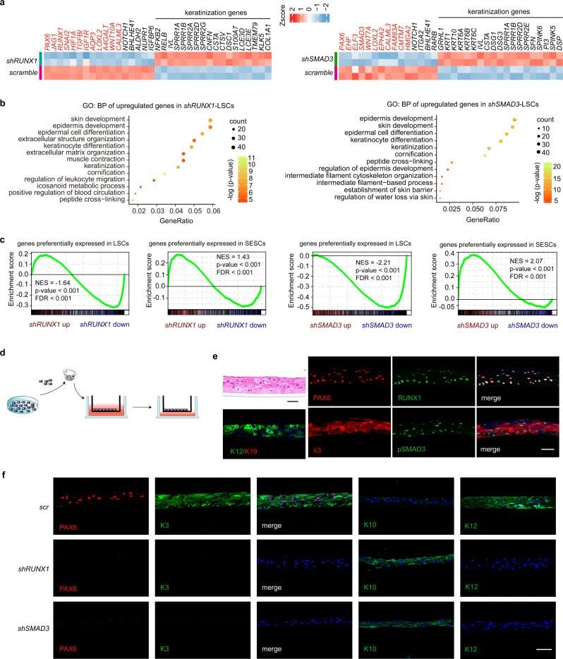
- Fig. 3 Loss of RUNX1 or SMAD3 induces cell identity switch. a Heatmaps of differentially expressed genes produced by RUNX1 or SMAD3 KD. Red mark represents SE-assigned genes. b GO BP analysis for the upregulated genes in RUNX1 -depleted and SMAD3 -depleted LSCs (pvalueCutoff = 0.01 and qvalueCutoff = 0.05). c GSEA for genes that (i) are expressed at higher levels in LSCs than in SESCs and (ii) are more highly expressed in SESCs than in LSCs. NES: normalized enrichment score. d Schema representation of the air-lifting culture system. LSCs were seeded in the transwell inserts and incubated in medium until they were confluent. Then, the medium in the upper chamber was removed to induce differentiation into a stratified epithelium sheet. e Hematoxylin and eosin (H&E) and immunofluorescence staining of the indicated genes in the differentiated corneal epithelium sheet after air-lifting induction. Scale bars, 50 mum. f Immunofluorescence staining of the indicated genes in the differentiated corneal epithelium sheets treated with the indicated shRNAs. Scale bar, 50 mum.
- Submitted by
- Invitrogen Antibodies (provider)
- Main image
- Experimental details
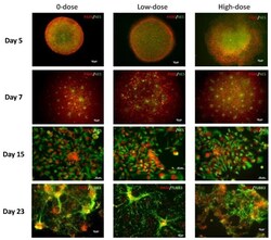
- Figure 2 Images of hESCs at different stages of differentiation to neuronal lineage. Staining of embryoid bodies (EBs) (Day 5); neural rosettes (Day 7); human neural progenitor cells (NPCs) (Day 15); and neural precursors (Day 23) with neuronal markers as indicated on the panels. Scale bar = 50 um.
