Explore
 Validate
 Learn
Learn Immunocytochemistry
Antibody data
- Antibody Data
- Antigen structure
- References [17]
- Comments [0]
- Validations
- Immunocytochemistry [1]
- Immunohistochemistry [3]
- Flow cytometry [1]
- Other assay [10]
Submit
Validation data
Reference
Comment
Report error
- Product number
- PA1-013 - Provider product page

- Provider
- Invitrogen Antibodies
- Product name
- KDEL Polyclonal Antibody
- Antibody type
- Polyclonal
- Antigen
- Synthetic peptide
- Description
- PA1-013 detects KDEL from human, rat, mouse and hamster.
- Concentration
- 1 mg/mL
Submitted references Posttranslational modifications of serine protease TMPRSS13 regulate zymogen activation, proteolytic activity, and cell surface localization.
Prohibitin 1 is essential to preserve mitochondria and myelin integrity in Schwann cells.
Dopamine Transporter Localization in Medial Forebrain Bundle Axons Indicates Its Long-Range Transport Primarily by Membrane Diffusion with a Limited Contribution of Vesicular Traffic on Retromer-Positive Compartments.
Induction of Axonal Outgrowth in Mouse Hippocampal Neurons via Bacterial Magnetosomes.
Extremely Low Forces Induce Extreme Axon Growth.
Ankyrin Is An Intracellular Tether for TMC Mechanotransduction Channels.
The Perinuclear ER Scales Nuclear Size Independently of Cell Size in Early Embryos.
TUBB1 mutations cause thyroid dysgenesis associated with abnormal platelet physiology.
CIB2 interacts with TMC1 and TMC2 and is essential for mechanotransduction in auditory hair cells.
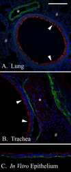
Hyaluronan Rafts on Airway Epithelial Cells.
Axons provide the secretory machinery for trafficking of voltage-gated sodium channels in peripheral nerve.
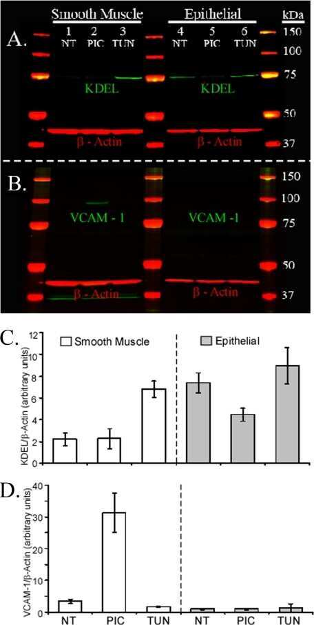
Primary murine airway smooth muscle cells exposed to poly(I,C) or tunicamycin synthesize a leukocyte-adhesive hyaluronan matrix.
Differentiated murine airway epithelial cells synthesize a leukocyte-adhesive hyaluronan matrix in response to endoplasmic reticulum stress.
Changes of endoplasmic reticulum chaperone complexes, redox state, and impaired protein disulfide reductase activity in misfolding alpha1-antitrypsin transgenic mice.
Further characterization of mammalian ceramide kinase: substrate delivery and (stereo)specificity, tissue distribution, and subcellular localization studies.
Functional proteomic screens reveal an essential extracellular role for hsp90 alpha in cancer cell invasiveness.
Protein-disulfide isomerase-mediated reduction of the A subunit of cholera toxin in a human intestinal cell line.
Martin CE, Murray AS, Sala-Hamrick KE, Mackinder JR, Harrison EC, Lundgren JG, Varela FA, List K
The Journal of biological chemistry 2021 Oct;297(4):101227
The Journal of biological chemistry 2021 Oct;297(4):101227
Prohibitin 1 is essential to preserve mitochondria and myelin integrity in Schwann cells.
Della-Flora Nunes G, Wilson ER, Marziali LN, Hurley E, Silvestri N, He B, O'Malley BW, Beirowski B, Poitelon Y, Wrabetz L, Feltri ML
Nature communications 2021 Jun 2;12(1):3285
Nature communications 2021 Jun 2;12(1):3285
Dopamine Transporter Localization in Medial Forebrain Bundle Axons Indicates Its Long-Range Transport Primarily by Membrane Diffusion with a Limited Contribution of Vesicular Traffic on Retromer-Positive Compartments.
Bagalkot TR, Block ER, Bucchin K, Balcita-Pedicino JJ, Calderon M, Sesack SR, Sorkin A
The Journal of neuroscience : the official journal of the Society for Neuroscience 2021 Jan 13;41(2):234-250
The Journal of neuroscience : the official journal of the Society for Neuroscience 2021 Jan 13;41(2):234-250
Induction of Axonal Outgrowth in Mouse Hippocampal Neurons via Bacterial Magnetosomes.
De Vincentiis S, Falconieri A, Mickoleit F, Cappello V, Schüler D, Raffa V
International journal of molecular sciences 2021 Apr 16;22(8)
International journal of molecular sciences 2021 Apr 16;22(8)
Extremely Low Forces Induce Extreme Axon Growth.
De Vincentiis S, Falconieri A, Mainardi M, Cappello V, Scribano V, Bizzarri R, Storti B, Dente L, Costa M, Raffa V
The Journal of neuroscience : the official journal of the Society for Neuroscience 2020 Jun 24;40(26):4997-5007
The Journal of neuroscience : the official journal of the Society for Neuroscience 2020 Jun 24;40(26):4997-5007
Ankyrin Is An Intracellular Tether for TMC Mechanotransduction Channels.
Tang YQ, Lee SA, Rahman M, Vanapalli SA, Lu H, Schafer WR
Neuron 2020 Jul 8;107(1):112-125.e10
Neuron 2020 Jul 8;107(1):112-125.e10
The Perinuclear ER Scales Nuclear Size Independently of Cell Size in Early Embryos.
Mukherjee RN, Sallé J, Dmitrieff S, Nelson KM, Oakey J, Minc N, Levy DL
Developmental cell 2020 Aug 10;54(3):395-409.e7
Developmental cell 2020 Aug 10;54(3):395-409.e7
TUBB1 mutations cause thyroid dysgenesis associated with abnormal platelet physiology.
Stoupa A, Adam F, Kariyawasam D, Strassel C, Gawade S, Szinnai G, Kauskot A, Lasne D, Janke C, Natarajan K, Schmitt A, Bole-Feysot C, Nitschke P, Léger J, Jabot-Hanin F, Tores F, Michel A, Munnich A, Besmond C, Scharfmann R, Lanza F, Borgel D, Polak M, Carré A
EMBO molecular medicine 2018 Dec;10(12)
EMBO molecular medicine 2018 Dec;10(12)
CIB2 interacts with TMC1 and TMC2 and is essential for mechanotransduction in auditory hair cells.
Giese APJ, Tang YQ, Sinha GP, Bowl MR, Goldring AC, Parker A, Freeman MJ, Brown SDM, Riazuddin S, Fettiplace R, Schafer WR, Frolenkov GI, Ahmed ZM
Nature communications 2017 Jun 29;8(1):43
Nature communications 2017 Jun 29;8(1):43
Hyaluronan Rafts on Airway Epithelial Cells.
Abbadi A, Lauer M, Swaidani S, Wang A, Hascall V
The Journal of biological chemistry 2016 Jan 15;291(3):1448-55
The Journal of biological chemistry 2016 Jan 15;291(3):1448-55
Axons provide the secretory machinery for trafficking of voltage-gated sodium channels in peripheral nerve.
González C, Cánovas J, Fresno J, Couve E, Court FA, Couve A
Proceedings of the National Academy of Sciences of the United States of America 2016 Feb 16;113(7):1823-8
Proceedings of the National Academy of Sciences of the United States of America 2016 Feb 16;113(7):1823-8
Primary murine airway smooth muscle cells exposed to poly(I,C) or tunicamycin synthesize a leukocyte-adhesive hyaluronan matrix.
Lauer ME, Mukhopadhyay D, Fulop C, de la Motte CA, Majors AK, Hascall VC
The Journal of biological chemistry 2009 Feb 20;284(8):5299-312
The Journal of biological chemistry 2009 Feb 20;284(8):5299-312
Differentiated murine airway epithelial cells synthesize a leukocyte-adhesive hyaluronan matrix in response to endoplasmic reticulum stress.
Lauer ME, Erzurum SC, Mukhopadhyay D, Vasanji A, Drazba J, Wang A, Fulop C, Hascall VC
The Journal of biological chemistry 2008 Sep 19;283(38):26283-96
The Journal of biological chemistry 2008 Sep 19;283(38):26283-96
Changes of endoplasmic reticulum chaperone complexes, redox state, and impaired protein disulfide reductase activity in misfolding alpha1-antitrypsin transgenic mice.
Papp E, Száraz P, Korcsmáros T, Csermely P
FASEB journal : official publication of the Federation of American Societies for Experimental Biology 2006 May;20(7):1018-20
FASEB journal : official publication of the Federation of American Societies for Experimental Biology 2006 May;20(7):1018-20
Further characterization of mammalian ceramide kinase: substrate delivery and (stereo)specificity, tissue distribution, and subcellular localization studies.
Van Overloop H, Gijsbers S, Van Veldhoven PP
Journal of lipid research 2006 Feb;47(2):268-83
Journal of lipid research 2006 Feb;47(2):268-83
Functional proteomic screens reveal an essential extracellular role for hsp90 alpha in cancer cell invasiveness.
Eustace BK, Sakurai T, Stewart JK, Yimlamai D, Unger C, Zehetmeier C, Lain B, Torella C, Henning SW, Beste G, Scroggins BT, Neckers L, Ilag LL, Jay DG
Nature cell biology 2004 Jun;6(6):507-14
Nature cell biology 2004 Jun;6(6):507-14
Protein-disulfide isomerase-mediated reduction of the A subunit of cholera toxin in a human intestinal cell line.
Orlandi PA
The Journal of biological chemistry 1997 Feb 14;272(7):4591-9
The Journal of biological chemistry 1997 Feb 14;272(7):4591-9
No comments: Submit comment
Supportive validation
- Submitted by
- Invitrogen Antibodies (provider)
- Main image
- Experimental details
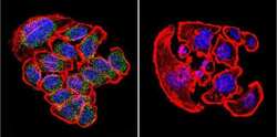
- Immunofluorescent analysis of KDEL in U251 Cells. Cells were grown on chamber slides and fixed with formaldehyde prior to staining. Cells were probed without (control) or with a KDEL polyclonal antibody (Product # PA1-013) at a dilution of 1:200 overnight at 4 C, washed with PBS and incubated with a DyLight-488 conjugated secondary antibody (Product # 35552). KDEL staining (green), F-Actin staining with Phalloidin (red) and nuclei with DAPI (blue) is shown. Images were taken at 60X magnification.
Supportive validation
- Submitted by
- Invitrogen Antibodies (provider)
- Main image
- Experimental details
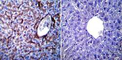
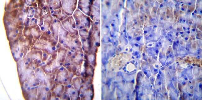
- Immunohistochemistry was performed on normal biopsies of deparaffinized Mouse liver tissue. To expose target proteins, heat induced antigen retrieval was performed using 10mM sodium citrate (pH6.0) buffer, microwaved for 8-15 minutes. Following antigen retrieval tissues were blocked in 3% BSA-PBS for 30 minutes at room temperature. Tissues were then probed at a dilution of 1:100 with a rabbit polyclonal antibody recognizing KDEL (Product # PA1-013) or without primary antibody (negative control) overnight at 4°C in a humidified chamber. Tissues were washed extensively with PBST and endogenous peroxidase activity was quenched with a peroxidase suppressor. Detection was performed using a biotin-conjugated secondary antibody and SA-HRP, followed by colorimetric detection using DAB. Tissues were counterstained with hematoxylin and prepped for mounting.

- Submitted by
- Invitrogen Antibodies (provider)
- Main image

- Experimental details
- Immunohistochemistry was performed on normal biopsies of deparaffinized Mouse lymph node tissue. To expose target proteins, heat induced antigen retrieval was performed using 10mM sodium citrate (pH6.0) buffer, microwaved for 8-15 minutes. Following antigen retrieval tissues were blocked in 3% BSA-PBS for 30 minutes at room temperature. Tissues were then probed at a dilution of 1:200 with a rabbit polyclonal antibody recognizing KDEL (Product # PA1-013) or without primary antibody (negative control) overnight at 4°C in a humidified chamber. Tissues were washed extensively with PBST and endogenous peroxidase activity was quenched with a peroxidase suppressor. Detection was performed using a biotin-conjugated secondary antibody and SA-HRP, followed by colorimetric detection using DAB. Tissues were counterstained with hematoxylin and prepped for mounting.

- Submitted by
- Invitrogen Antibodies (provider)
- Main image

- Experimental details
- Immunohistochemistry was performed on normal biopsies of deparaffinized Mouse pancreas tissue. To expose target proteins, heat induced antigen retrieval was performed using 10mM sodium citrate (pH6.0) buffer, microwaved for 8-15 minutes. Following antigen retrieval tissues were blocked in 3% BSA-PBS for 30 minutes at room temperature. Tissues were then probed at a dilution of 1:100 with a rabbit polyclonal antibody recognizing KDEL (Product # PA1-013) or without primary antibody (negative control) overnight at 4°C in a humidified chamber. Tissues were washed extensively with PBST and endogenous peroxidase activity was quenched with a peroxidase suppressor. Detection was performed using a biotin-conjugated secondary antibody and SA-HRP, followed by colorimetric detection using DAB. Tissues were counterstained with hematoxylin and prepped for mounting.
Supportive validation
- Submitted by
- Invitrogen Antibodies (provider)
- Main image
- Experimental details
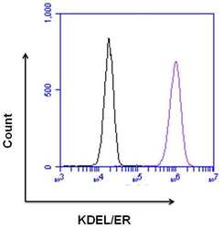
- Flow cytometry analysis of KDEL/ER was done on HeLa cells. Cells were fixed, permeabilized and stained with a KDEL/ER rabbit polyclonal antibody (Product # PA1-013, purple histogram) or a rabbit IgG isotype control (Product # MA5-16384, black histogram) at a dilution of 10 µg/mL. After incubation for 1 hour on ice, the cells were labeled with a Goat anti-Rabbit IgG (H+L) Superclonal™ Secondary Antibody, Alexa Fluor® 647 conjugate (Product # A27040) at a dilution of 1:50 for 1 hour on ice. A representative 10,000 cells were acquired and analyzed for each sample.

Supportive validation
- Submitted by
- Invitrogen Antibodies (provider)
- Main image
- Experimental details
- NULL
- Submitted by
- Invitrogen Antibodies (provider)
- Main image

- Experimental details
- NULL

- Submitted by
- Invitrogen Antibodies (provider)
- Main image

- Experimental details
- NULL

- Submitted by
- Invitrogen Antibodies (provider)
- Main image
- Experimental details
- NULL
- Submitted by
- Invitrogen Antibodies (provider)
- Main image

- Experimental details
- NULL

- Submitted by
- Invitrogen Antibodies (provider)
- Main image
- Experimental details
- NULL

- Submitted by
- Invitrogen Antibodies (provider)
- Main image
- Experimental details
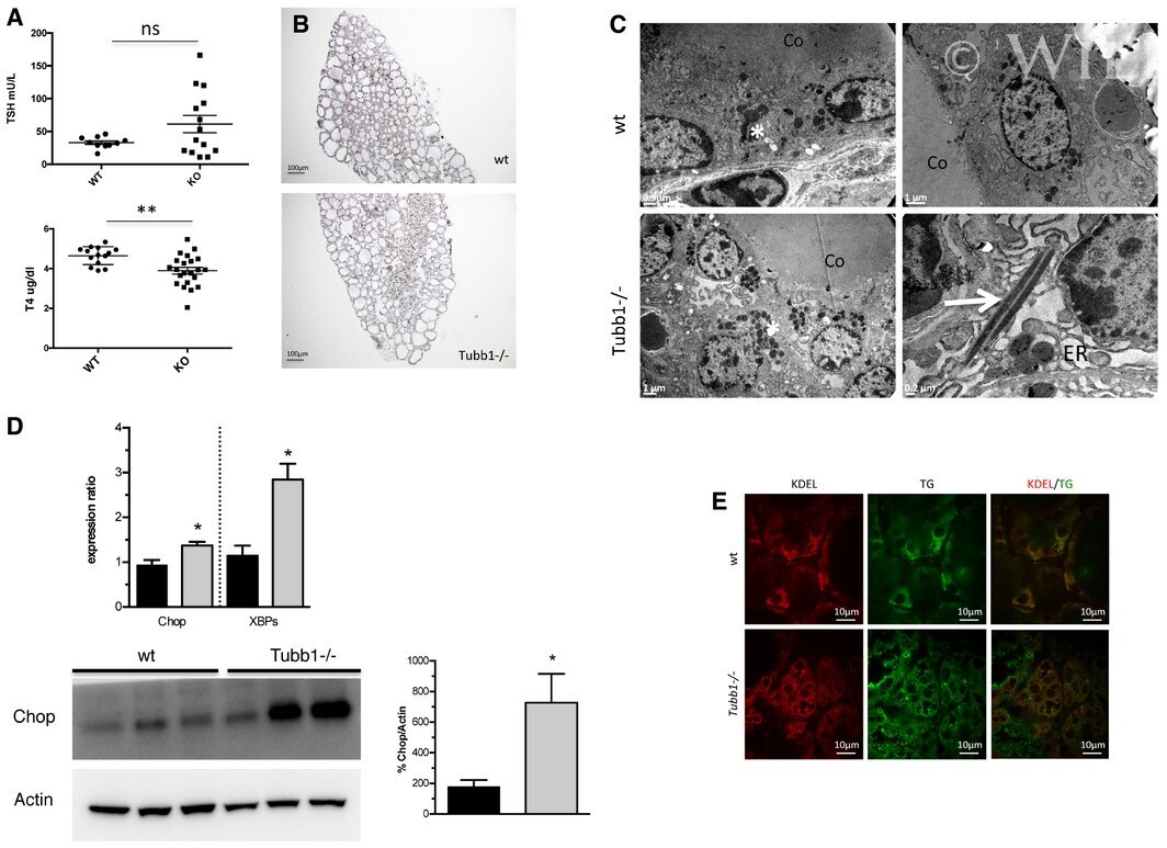
- Tubb1 -/- mice have altered thyroid function with impaired hormone secretion A Serum TSH and T4 levels in 3-month-old Tubb1 -/- and wild-type mice. Numbers of animals tested were 10 wild-type males and 14 Tubb1 -/- males for TSH and for 14 wild-type and 22 Tubb1 -/- males for T4. Tubb1 -/- mice had hypothyroidism with elevated TSH and decreased T4 versus wild-type mice. B Nkx2-1 (in brown) immunostaining of adult thyroid tissue. Note the disorganization of the thyroid tissue in Tubb1 -/- versus wild-type mice. C Ultrastructural alterations shown by electron microscopy in wild-type and Tubb1 -/- thyroid tissues. In Tubb1 -/- tissues, note the disorganization of secretion vesicles (white asterisks) and rods of identical density to secretion vesicles (white arrow). The ER is considerably dilated in Tubb1 -/- thyrocytes. Representative views. Scale bars at the bottom left for each view. Co: colloid. D ER stress in Tubb1 -/- thyroid tissue. Top: Chop and XBPs expression by quantitative PCR in adults, normalized to peptidylprolyl isomerase A. Experiments with four tissues per stage for each genotype; wt in black and Tubb1 -/- in grey. Bottom: Chop and Actin protein expression by Western blotting in three representative wild-type and Tubb1 -/- mice. The Chop band quantification normalized for Actin confirms the increased Chop expression demonstrated by quantitative PCR in Tubb1 -/- versus wild-type thyroids. All lanes are from the same blot, which
- Submitted by
- Invitrogen Antibodies (provider)
- Main image

- Experimental details
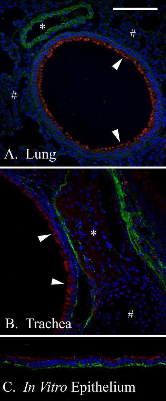
- Figure 3 Stretch-growth assay. ( A ) Length of primary axons in control (left) and stretched condition (right), scale bar: 50 um. ( B ) Length of primary axons of neurons treated with 1.25 ug Fe mL -1 of magnetosomes (box plot, min-to-max); n = 120 from four independent assays. T -test for unpaired data, p < 0.0001, t = 5.881, df = 238. ( C ) Length of primary axons of neurons treated with 2.5 ug Fe mL -1 of magnetosomes (box plot, min-to-max); n = 120 from four independent assays. T -test for unpaired data, p < 0.0001, t = 8.152, df = 238. ( D ) Axon caliber of neurons treated with 2.5 ug mL -1 of magnetosomes (scatter dot plot, mean +- SEM). T -test for unpaired data, n = 40 from four independent assays, p = 0.07, t = 1.817, df = 78. ( E ) Comparison between fold change of axon length in stretched versus non-stretched conditions of samples treated with 3.6 ug Fe mL -1 of SPIONs, with 1.25 ug mL -1 of magnetosomes (MS) and 2.5 ug mL -1 MS (box plot, min-to-max); one-way ANOVA test, n = 120 neurons from four independent assays, p = 0.021.

- Submitted by
- Invitrogen Antibodies (provider)
- Main image

- Experimental details
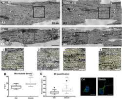
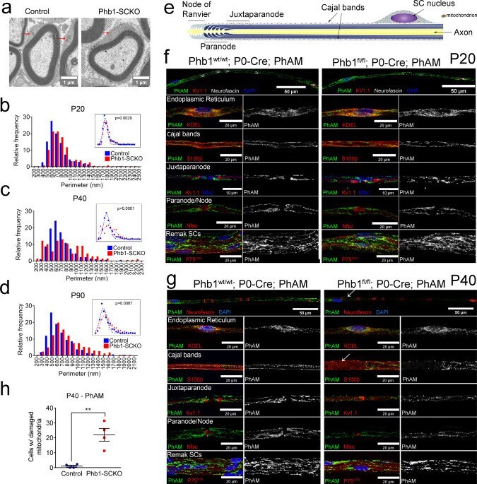
- Fig. 3 Ablation of Prohibitin 1 in Schwann cells results in altered mitochondrial morphology. a Representative electron micrographs highlighting the enlargement of mitochondria in sciatic nerves of Phb1-SCKO animals at postnatal day 20 (P20) (arrows). N = 3 animals per genotype. ( b - d ) Mitochondria in SCs of PHB1-SCKO mice (red) have a larger perimeter compared to mitochondria of control animals (blue) at P20 ( b ), P40 ( c ), and P90 ( d ). At P40, there is also a population of mitochondria that has a reduced perimeter, suggesting mitochondrial fragmentation. This population is lost at P90, suggesting that the fragmented mitochondria disappear. N = 3 animals per genotype; at least 100 mitochondria from each animal were evaluated. Insets: non-linear regression using a Gaussian curve followed by extra sum-of-squares F test [ F (3,42) P20 = 5.482, F (3,38) P40 = 19.48, F (3,34) P90 = 4.813). e Schematic representation of the distribution of mitochondria in a myelinating SC. f - g Confocal z-projections of teased fibers of sciatic nerves of Phb1-SCKO mice and controls illustrating the morphology of Schwann cell mitochondria as labeled by the PhAM reporter (green) near different cellular structures (red). DAPI is indicated in blue. f At P20, there are changes in mitochondrial size. N = 4 animals per genotype. g At P40, some cells lack PhAM expression away from the cell body. N = 3 animals per genotype. h PhAM is not detectable in about 20% of the myelin internodes of Phb1-SCKO
- Submitted by
- Invitrogen Antibodies (provider)
- Main image
- Experimental details
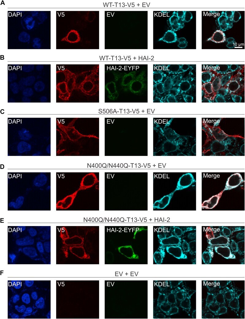
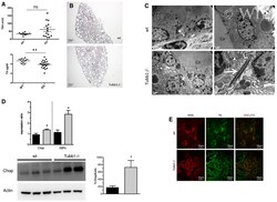
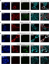
- Figure 4 Glycosylation deficiency causes ER retention of TMPRSS13. Twenty-four hours after seeding onto glass coverslips, HEK293T cells were transfected for 48 h with ( A ) WT-TMPRSS13 (T13)-V5 plus empty vector (EV), ( B ) WT-TMPRSS13-V5 plus HAI-2-EYFP, ( C ) S506A-TMPRSS13-V5 plus EV, ( D ) N400Q/N440Q-TMPRSS13-V5 plus EV, ( E ) N400Q/N440Q-TMPRSS13-V5 plus HAI-2-EYFP, or ( F ) EV plus EV. Cells were fixed, permeabilized, and incubated overnight with anti-V5 to detect TMPRSS13 and anti-KDEL to detect endogenous KDEL. Nuclei (DAPI) ( blue , A - F ), TMPRSS13-V5 ( red , A - F ), HAI-2 ( green , B and E ), KDEL ( cyan , A - F ). Merged images of TMPRSS13/KDEL are shown in panels on the right .