Explore
 Validate
 Learn
LearnMA5-13168
antibody from Invitrogen Antibodies
Targeting: MUC1
ADMCKD, ADMCKD1, CD227, MCD, MCKD, MCKD1, PEM, PUM
 Immunocytochemistry
 Immunohistochemistry
Antibody data
- Antibody Data
- Antigen structure
- References [12]
- Comments [0]
- Validations
- Immunohistochemistry [1]
- Flow cytometry [2]
- Other assay [4]
Submit
Validation data
Reference
Comment
Report error
- Product number
- MA5-13168 - Provider product page

- Provider
- Invitrogen Antibodies
- Product name
- Anti-MUC1 Monoclonal Antibody (GP1.4)
- Antibody type
- Monoclonal
- Antigen
- Other
- Description
- MA5-13168 targets Epithelial Membrane Antigen in IHC (P), ICC/IF and FACS applications and shows reactivity with human and mouse samples. The MA5-13168 immunogen is human milk fat globule membranes.
- Reactivity
- Human, Mouse
- Host
- Mouse
- Isotype
- IgG
- Antibody clone number
- GP1.4
- Vial size
- 500 µL
- Concentration
- 0.2 mg/mL
- Storage
- 4° C
Submitted references Mucin 1, a signal transduction membrane-bound mucin, is present in human disc tissue and is downregulated in vitro by exposure to IL-1ß or TNF-α.
Membrane-Tethered MUC1 Mucin Counter-Regulates the Phagocytic Activity of Macrophages.
Mucin1 shifts Smad3 signaling from the tumor-suppressive pSmad3C/p21(WAF1) pathway to the oncogenic pSmad3L/c-Myc pathway by activating JNK in human hepatocellular carcinoma cells.
Expression of human full-length MUC1 inhibits the proliferation and migration of a B16 mouse melanoma cell line.
Multinodular reticular schwannoma in the head and neck region: a potential diagnostic pitfall.
Anti-MUC1 antibody inhibits EGF receptor signaling in cancer cells.
Human embryonic stem cell-derived keratinocytes exhibit an epidermal transcription program and undergo epithelial morphogenesis in engineered tissue constructs.
Secondary involvement of the breast in T-cell non-Hodgkin lymphoma, an unusual example mimicking inflammatory breast carcinoma.
Myointimoma of the glans penis.
Pulmonary large cell carcinoma with rhabdoid phenotype.
Selective secretion and replenishment of discrete mucin glycoforms from intestinal goblet cells.
Selective secretion and replenishment of discrete mucin glycoforms from intestinal goblet cells.
Gruber HE, Ingram JA, Hoelscher GL, Marrero E, Hanley EN Jr
BMC musculoskeletal disorders 2017 May 8;18(1):182
BMC musculoskeletal disorders 2017 May 8;18(1):182
Membrane-Tethered MUC1 Mucin Counter-Regulates the Phagocytic Activity of Macrophages.
Kato K, Uchino R, Lillehoj EP, Knox K, Lin Y, Kim KC
American journal of respiratory cell and molecular biology 2016 Apr;54(4):515-23
American journal of respiratory cell and molecular biology 2016 Apr;54(4):515-23
Mucin1 shifts Smad3 signaling from the tumor-suppressive pSmad3C/p21(WAF1) pathway to the oncogenic pSmad3L/c-Myc pathway by activating JNK in human hepatocellular carcinoma cells.
Li Q, Liu G, Yuan H, Wang J, Guo Y, Chen T, Zhai R, Shao D, Ni W, Tai G
Oncotarget 2015 Feb 28;6(6):4253-65
Oncotarget 2015 Feb 28;6(6):4253-65
Expression of human full-length MUC1 inhibits the proliferation and migration of a B16 mouse melanoma cell line.
Wang F, Li Q, Ni W, Fang F, Sun X, Xie F, Wang J, Wang F, Gao S, Tai G
Oncology reports 2013 Jul;30(1):260-8
Oncology reports 2013 Jul;30(1):260-8
Multinodular reticular schwannoma in the head and neck region: a potential diagnostic pitfall.
Lau PP, Yau DT, Lau WH, Mak LS, Chan JK
International journal of surgical pathology 2013 Feb;21(1):54-8
International journal of surgical pathology 2013 Feb;21(1):54-8
Anti-MUC1 antibody inhibits EGF receptor signaling in cancer cells.
Hisatsune A, Nakayama H, Kawasaki M, Horie I, Miyata T, Isohama Y, Kim KC, Katsuki H
Biochemical and biophysical research communications 2011 Feb 18;405(3):377-81
Biochemical and biophysical research communications 2011 Feb 18;405(3):377-81
Human embryonic stem cell-derived keratinocytes exhibit an epidermal transcription program and undergo epithelial morphogenesis in engineered tissue constructs.
Metallo CM, Azarin SM, Moses LE, Ji L, de Pablo JJ, Palecek SP
Tissue engineering. Part A 2010 Jan;16(1):213-23
Tissue engineering. Part A 2010 Jan;16(1):213-23
Secondary involvement of the breast in T-cell non-Hodgkin lymphoma, an unusual example mimicking inflammatory breast carcinoma.
Kelten C, Kabukcu S, Sen N, Teke Z, Yaren A, Erdem E, Duzcan E
Archives of gynecology and obstetrics 2009 Jul;280(1):149-52
Archives of gynecology and obstetrics 2009 Jul;280(1):149-52
Myointimoma of the glans penis.
Vardar E, Gunlusoy B, Arslan M, Kececi S
Pathology international 2007 Mar;57(3):158-61
Pathology international 2007 Mar;57(3):158-61
Pulmonary large cell carcinoma with rhabdoid phenotype.
Yilmazbayhan D, Ates LE, Dilege S, Gulluoglu M, Tanju S, Kalayci G
Annals of diagnostic pathology 2005 Aug;9(4):223-6
Annals of diagnostic pathology 2005 Aug;9(4):223-6
Selective secretion and replenishment of discrete mucin glycoforms from intestinal goblet cells.
Stanley CM, Phillips TE
The American journal of physiology 1999 Jul;277(1):G191-200
The American journal of physiology 1999 Jul;277(1):G191-200
Selective secretion and replenishment of discrete mucin glycoforms from intestinal goblet cells.
Stanley CM, Phillips TE
The American journal of physiology 1999 Jul;277(1 Pt 1):G191-200
The American journal of physiology 1999 Jul;277(1 Pt 1):G191-200
No comments: Submit comment
Supportive validation
- Submitted by
- Invitrogen Antibodies (provider)
- Main image

- Experimental details
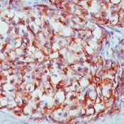
- Formalin-fixed, paraffin-embedded human breast carcinoma stained with EMA antibody using peroxidase-conjugate and AEC chromogen. Note membrane staining of tumor cells.
Supportive validation
- Submitted by
- Invitrogen Antibodies (provider)
- Main image

- Experimental details
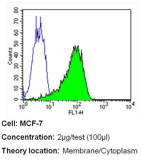
- Flow cytometry analysis of Mucin 1 in MCF-7 cells (green) compared to an isotype control (blue). Cells were harvested, adjusted to a concentration of 1-5x10^6 cells/mL, fixed with 2% paraformaldehyde and washed with PBS. Cells were blocked with a 2% solution of BSA-PBS for 30 min at room temperature and incubated with a Mucin 1 monoclonal antibody (Product # MA5-13168) at a dilution of 2 µg/test for 40 min at room temperature. Cells were then incubated for 40 min at room temperature in the dark using a Dylight 488-conjugated secondary antibody and re-suspended in PBS for FACS analysis.
- Submitted by
- Invitrogen Antibodies (provider)
- Main image
- Experimental details
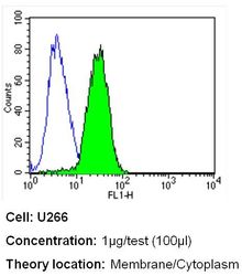
- Flow cytometry analysis of Mucin 1 in U266 cells (green) compared to an isotype control (blue). Cells were harvested, adjusted to a concentration of 1-5x10^6 cells/mL, fixed with 2% paraformaldehyde and washed with PBS. Cells were blocked with a 2% solution of BSA-PBS for 30 min at room temperature and incubated with a Mucin 1 monoclonal antibody (Product # MA5-13168) at a dilution of 1 µg/test for 40 min at room temperature. Cells were then incubated for 40 min at room temperature in the dark using a Dylight 488-conjugated secondary antibody and re-suspended in PBS for FACS analysis.

Supportive validation
- Submitted by
- Invitrogen Antibodies (provider)
- Main image
- Experimental details
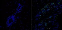
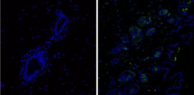
- Immunofluorescent analysis of Mucin 1 (green) showing staining in the membrane of mouse breast tissue (right) compared to a negative control without primary antibody (left). Formalin-fixed tissue was permeabilized with 0.1% Triton X-100 in TBS for 5-10 minutes and blocked with 3% BSA-PBS for 30 minutes at room temperature. Tissue was probed with a Mucin 1 monoclonal antibody (Product # MA5-13168) in 3% BSA-PBS at a dilution of 1:50 and incubated overnight at 4ºC in a humidified chamber. Tissue was washed with PBST and incubated with a DyLight-conjugated secondary antibody in PBS at room temperature in the dark. Nuclei were stained with Hoechst or DAPI (blue). Images were taken at a magnification of 60x.
- Submitted by
- Invitrogen Antibodies (provider)
- Main image
- Experimental details
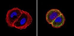
- Immunofluorescent analysis of Mucin 1 (green) showing staining in the cytoplasm and membrane of MCF-7 cells (right) compared to a negative control without primary antibody (left). Formalin-fixed cells were permeabilized with 0.1% Triton X-100 in TBS for 5-10 minutes and blocked with 3% BSA-PBS for 30 minutes at room temperature. Cells were probed with a Mucin 1 monoclonal antibody (Product # MA5-13168) in 3% BSA-PBS at a dilution of 1:50 and incubated overnight at 4ºC in a humidified chamber. Cells were washed with PBST and incubated with a DyLight-conjugated secondary antibody in PBS at room temperature in the dark. Actin was stained using Alexa Fluor 554 (red) and nuclei were stained with Hoechst or DAPI (blue). Images were taken at a magnification of 60x.

- Submitted by
- Invitrogen Antibodies (provider)
- Main image
- Experimental details
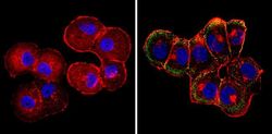
- Immunofluorescent analysis of Mucin 1 (green) showing staining in the cytoplasm and membrane of T47D cells (right) compared to a negative control without primary antibody (left). Formalin-fixed cells were permeabilized with 0.1% Triton X-100 in TBS for 5-10 minutes and blocked with 3% BSA-PBS for 30 minutes at room temperature. Cells were probed with a Mucin 1 monoclonal antibody (Product # MA5-13168) in 3% BSA-PBS at a dilution of 1:100 and incubated overnight at 4ºC in a humidified chamber. Cells were washed with PBST and incubated with a DyLight-conjugated secondary antibody in PBS at room temperature in the dark. Actin was stained using Alexa Fluor 554 (red) and nuclei were stained with Hoechst or DAPI (blue). Images were taken at a magnification of 60x.

- Submitted by
- Invitrogen Antibodies (provider)
- Main image
- Experimental details
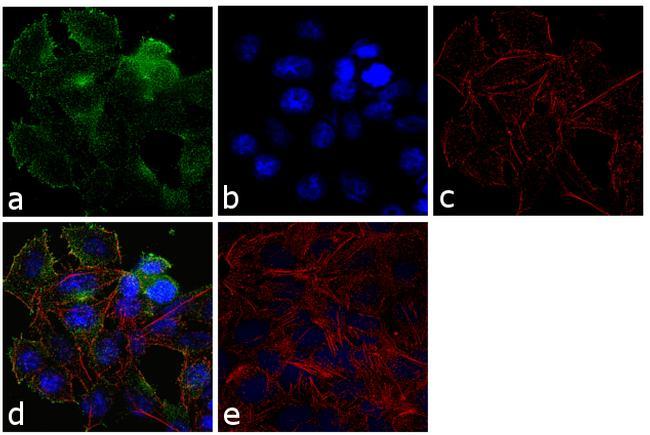
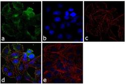
- Immunofluorescence analysis of MUC1 was performed using 70% confluent log phase MCF7 cells. The cells were fixed with 4% paraformaldehyde for 10 minutes, permeabilized with 0.1% Triton™ X-100 for 10 minutes, and blocked with 1% BSA for 1 hour at room temperature. The cells were labeled with MUC1 (GP1.4) Mouse Monoclonal Antibody (Product # MA5-13168) at 2µg/mL in 0.1% BSA and incubated for 3 hours at room temperature and then labeled with Goat anti-Mouse IgG (H+L) Superclonal™ Secondary Antibody, Alexa Fluor® 488 conjugate (Product # A28175) at a dilution of 1:2000 for 45 minutes at room temperature (Panel a: green). Nuclei (Panel b: blue) were stained with SlowFade® Gold Antifade Mountant with DAPI (Product # S36938). F-actin (Panel c: red) was stained with Rhodamine Phalloidin (Product # R415, 1:300). Panel d represents the merged image showing membranous and cytoplasmic localization. Panel e shows the no primary antibody control. The images were captured at 60X magnification.