Explore
 Validate
 Learn
Learn Western blot
Antibody data
- Antibody Data
- Antigen structure
- References [0]
- Comments [0]
- Validations
- Western blot [2]
- Immunocytochemistry [2]
Submit
Validation data
Reference
Comment
Report error
- Product number
- MA3-089 - Provider product page

- Provider
- Invitrogen Antibodies
- Product name
- FUS Monoclonal Antibody (1FU-1D2)
- Antibody type
- Monoclonal
- Antigen
- Synthetic peptide
- Description
- MA3-089 detects AID from human samples
- Antibody clone number
- 1FU-1D2
- Concentration
- Conc. Not Determined
No comments: Submit comment
Supportive validation
- Submitted by
- Invitrogen Antibodies (provider)
- Main image
- Experimental details
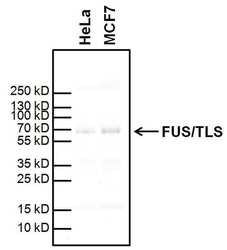
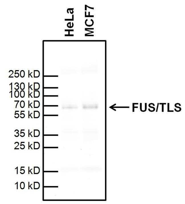
- Western blot analysis of FUS/TLS was performed by loading 10 µg of acid extracted nuclear lysate from HeLa cells (lane 1) and MCF7 cells (lane 2) in reducing sample buffer (Product # 39000) and Page Ruler Plus Protein Ladder (Product # 26619) onto a Novex 4-20% Tris-Glycine polyacrylamide gel (Product # WT4201BX10). Proteins were transferred to nitrocellulose membrane (Product # 88018) with Transfer Buffer (Product # 84731) using the G2 Fast Blotter (Product # 62288). Membrane was blocked in StartingBlock T20 (Product # 37543) for 30 min at room temperature. FUS/TLS was detected at approximately 70 kDa using a FUS/TLS monoclonal antibody (Product # MA3-089) at a dilution of 1:2000 in StartingBlock T20 overnight at at 4°C on a rocking platform, followed by a goat anti-mouse superclonal IgG-HRP secondary antibody (Product # A28177) at a dilution of 1:5000 for one hour. Chemiluminescent detection was performed using SuperSignal West Pico (Product # 34078) and the myECL Imager (Product # 62236).
- Submitted by
- Invitrogen Antibodies (provider)
- Main image
- Experimental details
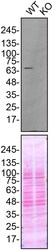
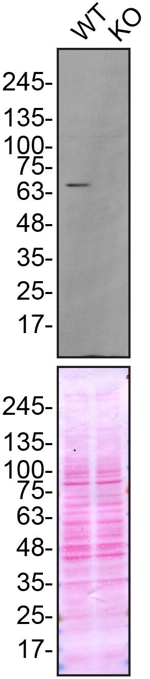
- Western blot of FUS was performed by loading 30 µg of WT (lane 1) and FUS CRISPR KO (lane 2) HeLa cell lysates in RIPA buffer onto a 5-16% gradient polyacrylamide gel. Proteins on the blots were visualized with Ponceau staining (below immunoblot). Proteins were transferred to nitrocellulose membrane and blocked in 5% milk for 1 hr. FUS was detected at approximately 70 kDa using a FUS monoclonal antibody (Product # MA3-089) at a dilution of 1:5,000 in 5% BSA in TBS with 0.1% Tween 20 (TBST) overnight at 4°C. The peroxidase-conjugated secondary antibody (Product # 62-6520) was diluted to 0.2 µg/mL in TBST with 5% milk for 1 hr. Chemiluminescent detection was performed using Pierce ECL Western Blotting Substrate (Product # 32106). Data courtesy of YCharOS Inc., an open science company with the mission of characterizing commercially available antibodies using knockout validation.
Supportive validation
- Submitted by
- Invitrogen Antibodies (provider)
- Main image
- Experimental details
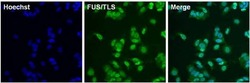
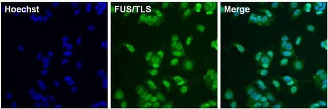
- Immunofluorescent analysis of FUS/TLS (green) in MCF7 cells. The cells were fixed with 4% formaldehyde (Product # 28908) for 15 min, permeabilized with 1X Permeablilization buffer (Product # 8408400) for 15 minutes, and blocked with 1% Blocker BSA (Product # 37525) for 15 minutes at room temperature. Cells were stained with FUS/TLS monoclonal antibody (Product # MA3-089) at a dilution of 1:500 in blocking buffer overnight at room temperature, washed with 1X TBS Tween 20 Buffer (Product # 28360), followed by incubation with DyLight 488 goat anti-mouse IgG secondary antibody (green, Product # A28175) at a dilution of 1:500 and Hoechst 33342 dye (blue, Product # 62249) at a dilution of 1:5000 for 30 minutes at room temperature. Images were taken on a Thermo Scientific ToxInsight at 20X magnification.
- Submitted by
- Invitrogen Antibodies (provider)
- Main image
- Experimental details
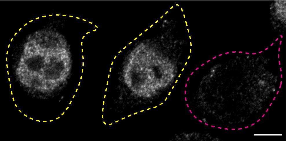
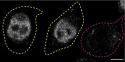
- Immunofluorescence of FUS was performed using HeLa wild-type and FUS KO cells that were transfected with a green or a far red fluorescent dye, respectively. Post-transfection, WT and KO cells were mixed and plated to a 1:1 ratio on coverslips as a mosaic and incubated for 24 hrs. Cells were fixed in 4% PFA (in PBS) for 15 min; cells were permeabilized with 0.1% Triton X-100 for 10 min at RT and blocked with PBS with 5% BSA, 5% goat serum, and 0.01% Triton X-100 for 30 min. Cells were stained with the FUS monoclonal antibody (Product # MA3-089) at a 1:1,000 dilution overnight at 4°C. Secondary antibody incubation was performed using 1 µg/mL of Goat anti-Mouse IgG (H+L) Highly Cross-Adsorbed Secondary Antibody, Alexa Fluor 555 antibody (Product # A21424) for 1 hr. Imaging was performed with a 40X oil objective and analysis was performed using Image J. Cell image represents a single focal plane; WT and KO cells are outlined with a yellow (WT) or magenta (KO) dashed line. Data courtesy of YCharOS Inc., an open science company with the mission of characterizing commercially available antibodies using knockout validation.