Explore
 Validate
 Learn
Learn Western blot
Antibody data
- Antibody Data
- Antigen structure
- References [1]
- Comments [0]
- Validations
- Western blot [3]
- Immunocytochemistry [1]
- Immunohistochemistry [5]
- Other assay [9]
Submit
Validation data
Reference
Comment
Report error
- Product number
- PA5-52610 - Provider product page

- Provider
- Invitrogen Antibodies
- Product name
- FUS Polyclonal Antibody
- Antibody type
- Polyclonal
- Antigen
- Recombinant full-length protein
- Description
- Immunogen sequence: SSQSSYGQQS SYPGYGQQPA PSSTSGSYGS SSQSSSYGQP QSGSYSQQPS YGGQQQSYGQ QQSYNPPQGY GQQNQYNSSS GGGGGGGGGG NYGQDQSSMS SGGGSGGGYG NQDQSGGGGS GGYGQQDR
- Concentration
- 0.09 mg/mL
Submitted references FUS driven circCNOT6L biogenesis in mouse and human spermatozoa supports zygote development.
Chioccarelli T, Falco G, Cappetta D, De Angelis A, Roberto L, Addeo M, Ragusa M, Barbagallo D, Berrino L, Purrello M, Ambrosino C, Cobellis G, Pierantoni R, Chianese R, Manfrevola F
Cellular and molecular life sciences : CMLS 2021 Dec 22;79(1):50
Cellular and molecular life sciences : CMLS 2021 Dec 22;79(1):50
No comments: Submit comment
Supportive validation
- Submitted by
- Invitrogen Antibodies (provider)
- Main image
- Experimental details
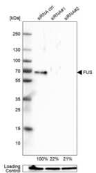
- Western blot analysis of FUS in U-251MG cells transfected with control siRNA, target specific siRNA probe #1 and #2, using a FUS Polyclonal Antibody (Product # PA5-52610). Remaining relative intensity is presented. Loading control: Anti-GAPDH.
- Submitted by
- Invitrogen Antibodies (provider)
- Main image
- Experimental details
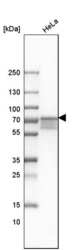
- Western blot analysis of FUS in human cell line HeLa using a FUS Polyclonal Antibody (Product # PA5-52610).

- Submitted by
- Invitrogen Antibodies (provider)
- Main image

- Experimental details
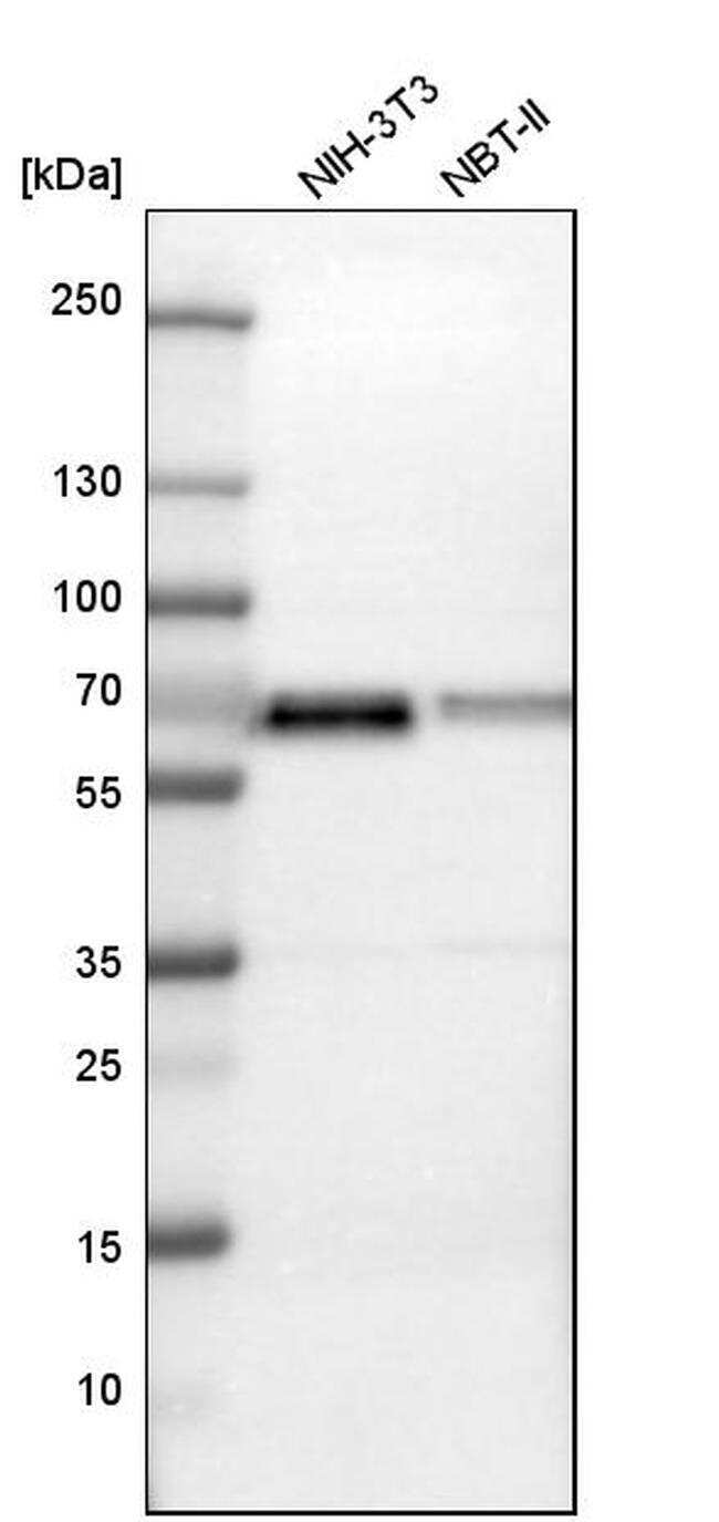
- Western blot analysis of FUS in mouse cell line NIH-3T3 and rat cell line NBT-II using a FUS Polyclonal Antibody (Product # PA5-52610).
Supportive validation
- Submitted by
- Invitrogen Antibodies (provider)
- Main image

- Experimental details
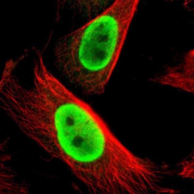
- Immunofluorescent staining of FUS in human cell line U-251 MG shows positivity in nucleus but excluded from the nucleoli. Samples were probed using a FUS Polyclonal Antibody (Product # PA5-52610).
Supportive validation
- Submitted by
- Invitrogen Antibodies (provider)
- Main image
- Experimental details
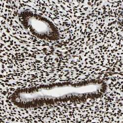
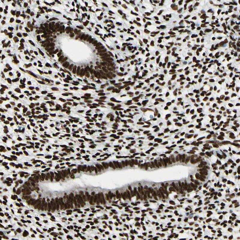
- Immunohistochemical staining of FUS in human endometrium using a FUS Polyclonal Antibody (Product # PA5-52610) shows strong nuclear positivity in glandular cells and stromal cells.
- Submitted by
- Invitrogen Antibodies (provider)
- Main image

- Experimental details
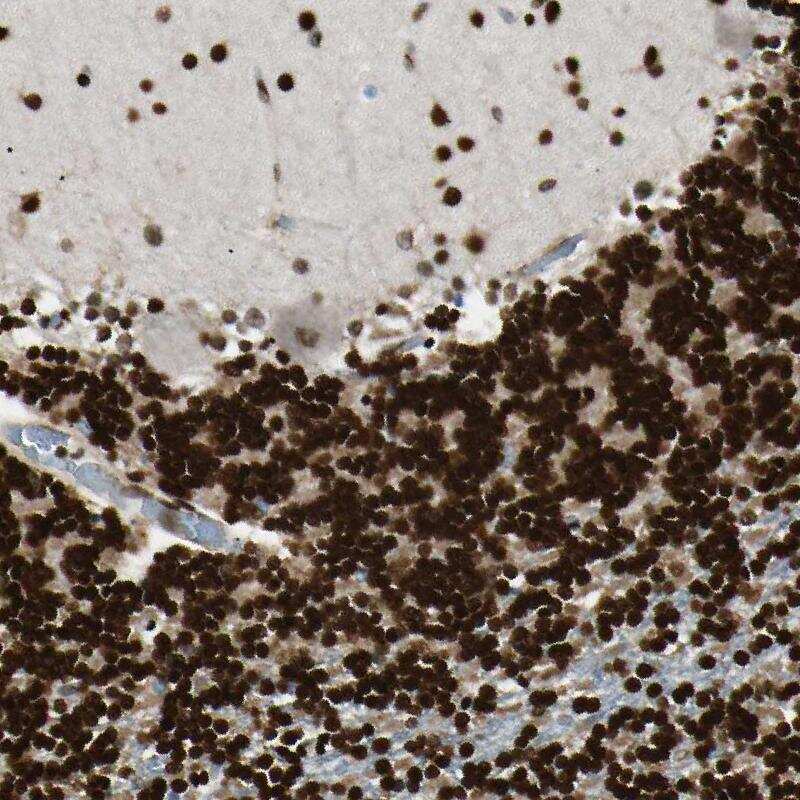
- Immunohistochemical staining of FUS in human testis using a FUS Polyclonal Antibody (Product # PA5-52610) shows strong nuclear positivity in cells in seminiferous ducts.

- Submitted by
- Invitrogen Antibodies (provider)
- Main image
- Experimental details
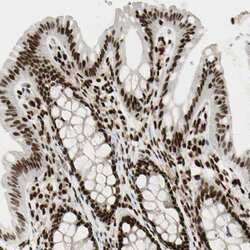
- Immunohistochemical staining of FUS in human colon using a FUS Polyclonal Antibody (Product # PA5-52610) shows strong nuclear positivity in glandular cells.

- Submitted by
- Invitrogen Antibodies (provider)
- Main image
- Experimental details
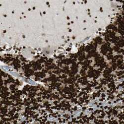
- Immunohistochemical staining of FUS in human cerebellum using a FUS Polyclonal Antibody (Product # PA5-52610) shows strong nuclear positivity in neuronal cells.
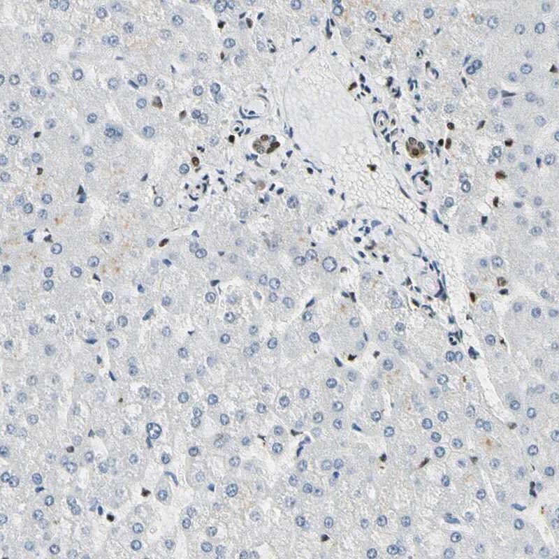
- Submitted by
- Invitrogen Antibodies (provider)
- Main image
- Experimental details
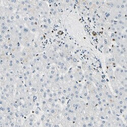
- Immunohistochemical staining of FUS in human liver using FUS Polyclonal Antibody (Product # PA5-52610) shows moderate nuclear positivity in bile duct cells while hepatocytes are negative.
Supportive validation
- Submitted by
- Invitrogen Antibodies (provider)
- Main image
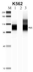
- Experimental details
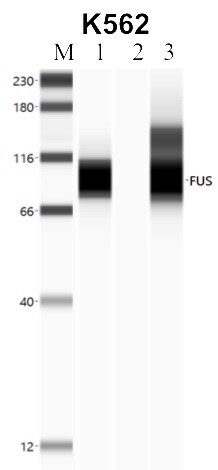
- RNA immunoprecipitation (RIP) western of FUS was performed in K562 cells. Antigen-antibody complexes were formed by incubating approximately 500 µg whole cell lysate with 5 to 10 µL of polyclonal FUS antibody (Product # PA5-52610) rotating 60 min at RT. The immune complexes were captured on 625 µg of anti- rabbit coated Dynabeads (Product # 11204D) and washed extensively. They were then eluted and analyzed using the Simple Western system using the same antibody as used in immunoprecipitation at a dilution of 1:25, followed by a 1:100 dilution of secondary antibody. Lane 1 is the input, lane 2 no antibody IP and lane 3 is the target specific IP. Data courtesy of the Yeo lab as part of the ENCODE project.
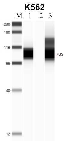
- Submitted by
- Invitrogen Antibodies (provider)
- Main image
- Experimental details
- RNA immunoprecipitation (RIP) western of FUS was performed in K562 cells. Antigen-antibody complexes were formed by incubating approximately 500 µg whole cell lysate with 5 to 10 µL of polyclonal FUS antibody (Product # PA5-52610) rotating 60 min at RT. The immune complexes were captured on 625 µg of anti- rabbit coated Dynabeads (Product # 11204D) and washed extensively. They were then eluted and analyzed using the Simple Western system using the same antibody as used in immunoprecipitation at a dilution of 1:25, followed by a 1:100 dilution of secondary antibody. Lane 1 is the input, lane 2 no antibody IP and lane 3 is the target specific IP. Data courtesy of the Yeo lab as part of the ENCODE project.
- Submitted by
- Invitrogen Antibodies (provider)
- Main image
- Experimental details
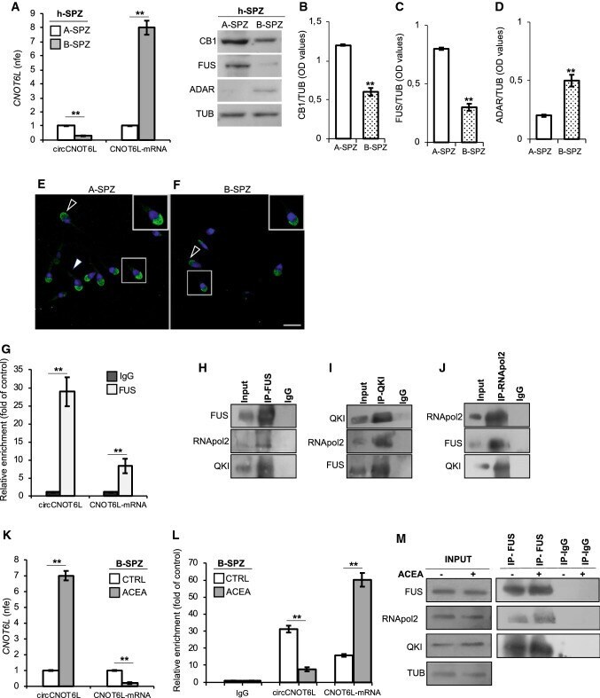
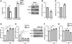
- Fig. 1 CB1 involvement in circCNOT6L biogenesis in testis. qRT-PCR detection of circCNOT6L and CNOT6L-mRNA expression levels ( A ); immunoblots and quantification of FUS ( B ) and ADAR ( C ) proteins in testis of WT and Cb1 -/- adult mice ( n = 3 mice in triplicate for each group). qRT-PCR detection of circCNOT6L ( D ), CNOT6L-mRNA ( E ) expression levels and immunoblots and quantification of FUS ( F ) and ADAR ( G ) proteins in testis of WT mice in vitro treated with vehicle (CTRL) or ACEA at different concentrations: 0, 1-1-10 uM ( n = 3 different testes in triplicate for each experimental group). In ( A ), ( D ) and ( E ), data are reported as mean value of nfe +- SEM, using Actin as endogenous control, while in ( B ), ( C ), ( F ) and ( G ), FUS and ADAR amount was quantified by densitometry analysis, normalized against Tubulin (TUB) signals, expressed as fc of OD values and reported as mean value +- SEM. ** P < 0.01

- Submitted by
- Invitrogen Antibodies (provider)
- Main image

- Experimental details
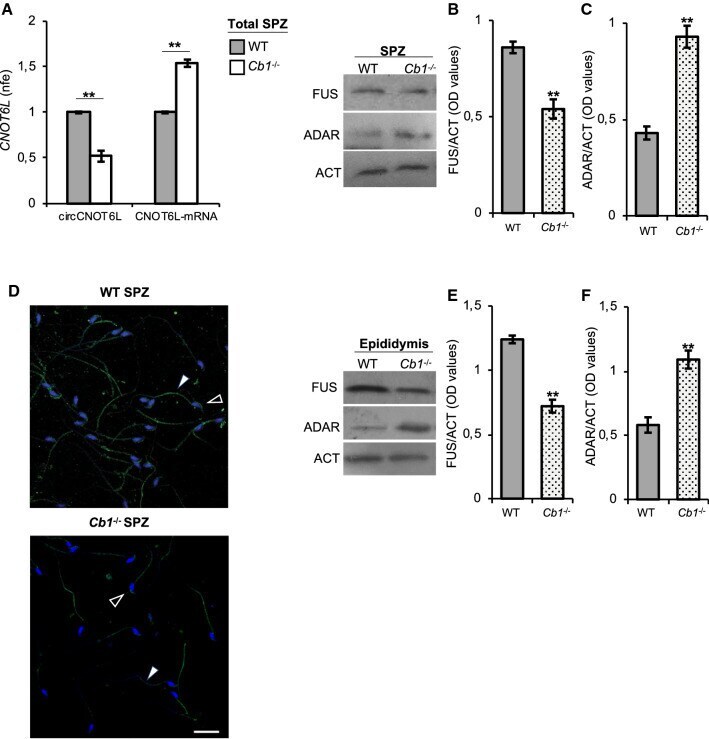
- Fig. 2 CB1 involvement in circCNOT6L biogenesis in total epididymal SPZ. qRT-PCR detection of circCNOT6L and CNOT6L-mRNA expression levels ( A ); immunoblots and quantification of FUS ( B and E ) and ADAR ( C and F ) proteins in total epididymal SPZ and/or epididymal tissue from WT and Cb1 -/- adult mice ( n = 3 mice in triplicate for each group). In ( A ), the data are reported as mean value of nfe +- SEM, using Actin as endogenous control, while in ( B ), ( C ), ( E ) and ( F ), FUS and ADAR amount was quantified by densitometry analysis, normalized against Actin (ACT) signals, expressed as OD values and reported as mean value +- SEM. ** P < 0.01. Immunofluorescence analysis of FUS protein in WT and Cb1 -/- SPZ ( D ). White empty arrowheads and white full arrowheads represent FUS localization (FITC-green) in sperm head and tail, respectively. Nuclei were labeled with DAPI (blue). Scale bar: 20 uM
- Submitted by
- Invitrogen Antibodies (provider)
- Main image
- Experimental details
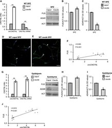
- Fig. 3 Differential analysis of circCNOT6L in WT caput and cauda SPZ and epididymal tissue. qRT-PCR detection of circCNOT6L and CNOT6L-mRNA expression levels ( A and G ); immunoblots and quantification of FUS ( B and H ) and ADAR ( C and I ) proteins in caput and cauda SPZ ( A , B and C ) and epididymal tissue ( G , H and I ) from WT adult mice ( n = 3 different samples from three different mice in triplicate). In ( A ) and ( G ) data are reported as mean value of nfe +- SEM, using Actin as endogenous control, while in ( B ), ( C ), ( H ) and ( I ), FUS and ADAR amount was quantified by densitometry analysis, normalized against Actin (ACT) signals, expressed as fc of OD values and reported as mean value +- SEM. * P < 0.05, ** P < 0.01. Immunofluorescence analysis of FUS protein in WT caput and cauda SPZ ( D and E ). White empty arrowheads and white full arrowheads represent FUS localization (FITC-green) in sperm head and tail, respectively. Nuclei were labeled with DAPI (blue). Scale bar: 20 uM. Correlation analysis ( F and J ) between circCNOT6L and FUS expression values relative to caput and cauda SPZ ( F ; r = 0.905, P < 0.001) and epididymal tissue ( J ; r = 0.910, P < 0.001) of WT mice regardless of the epididymal region

- Submitted by
- Invitrogen Antibodies (provider)
- Main image

- Experimental details
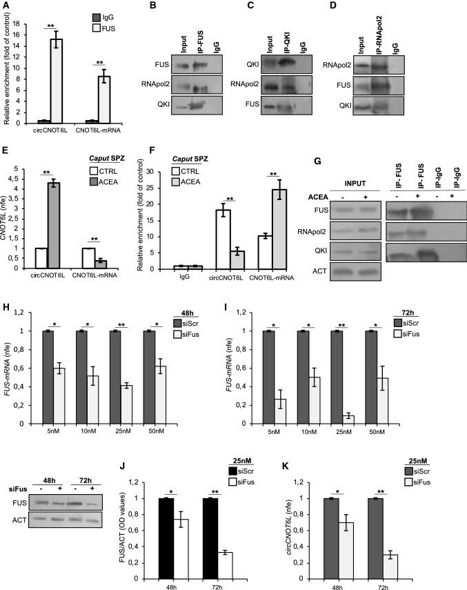
- Fig. 4 FUS drives CNOT6L backsplicing by interacting with RNApol2 and QKI in caput SPZ. The enrichment levels of circCNOT6L and CNOT6L-mRNA in the products of RIP assay (FUS-IP compared with IgG-IP) in WT caput SPZ alone ( A ) and after in vitro ACEA treatment ( F ) detected by qRT-PCR. Data are reported as mean +- SEM from three independent experiments. ** P < 0.01. qRT-PCR detection of circCNOT6L and CNOT6L-mRNA expression levels ( E ) in caput SPZ from WT mice in vitro treated with vehicle (CTRL) or ACEA 1 uM ( n = 3 different samples from three different animals for each experimental group in triplicate). Data are reported as mean value of nfe +- SEM, using Actin as endogenous control. ** P < 0.01. Western blot analysis of FUS, QKI and RNApol2 in the products of IP in WT caput SPZ ( B , C and D ) using FUS, QKI and RNApol2 antibodies. Western blot analysis of RIP protein fraction immunoprecipitated with FUS Ab (FUS-IP) in WT caput SPZ after in vitro ACEA treatment ( G ). FUS-IP is analyzed in comparison to control IgG-IP and Input protein extracts. qRT-PCR detection of Fus expression levels in ESCs, cultured in RM medium, treated with siScr or siFus (5, 10, 25 and 50 nM) and harvested after 48 ( H ) and 72 h ( I ) from siRNA transfection. Data are reported as mean value of nfe +- SEM, using Actin as endogenous control. ** P < 0.01; * P < 0.05. Western blot analysis of FUS protein ( J ) and qRT-PCR detection of circCNOT6L expression levels ( K ) in ESCs, cultured in RM med
- Submitted by
- Invitrogen Antibodies (provider)
- Main image
- Experimental details
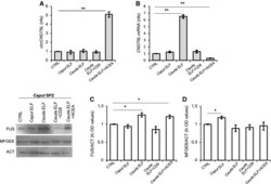
- Fig. 5 In vitro experiment of vesicle shuttle: from epididymis to SPZ. qRT-PCR detection of circCNOT6L ( A ) and CNOT6L-mRNA ( B ) expression levels in caput SPZ from WT mice in vitro co-incubated with: PBS (CTRL group), Caput ELF, Cauda ELF, Cauda ELF pre-treated with anti-CD9 Ab ( Cauda ELF + CD9), Cauda ELF in presence of ACEA 1 muM ( Cauda ELF + ACEA); ( n = 3 different samples for each experimental group from 8 different animals in triplicate). Data are reported as mean value nfe +- SEM, using Actin as endogenous control. ** P < 0.01. Immunoblots and quantification of FUS ( C ) and MFG-E8 ( D ) proteins in caput SPZ from WT mice in vitro co-incubated with: PBS (CTRL group), Caput ELF, Cauda ELF, Cauda ELF pre-treated with anti-CD9 Ab ( Cauda ELF + CD9), Cauda ELF in presence of ACEA 1 muM ( Cauda ELF + ACEA); ( n = 3 different samples for each experimental group from eight different animals in triplicate). FUS and MFG-E8 amount was quantified by densitometry analysis, normalized against Actin (ACT) signals and expressed as fc of OD values. Data are reported as mean value of fc of OD values +- SEM. * P < 0.05

- Submitted by
- Invitrogen Antibodies (provider)
- Main image
- Experimental details
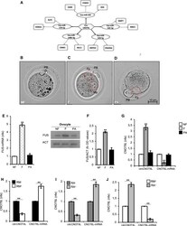
- Fig. 6 circCNOT6L from SPZ to oocyte toward the 2-cell-like state. CircCNOT6L-miRNA-mRNA network analysis was carried out by using the bioinformatic online programs (starBase, circBase, TargetScan, miRBase, Cytoscape) ( A ). Representative image of NF, F and PA ( B , C and D ). Fp female pronucleus, Mp male pronucleus, PB first polar globule. Scale bar: 10 uM. Expression analysis of FUS mRNA ( E ) and immunoblots and quantification of FUS protein ( F ) in NF, F and PA. n = 3 pools each containing 10 NF, n = 3 pools each containing 10 F and n = 3 pools each containing 10 PA were used for expression analysis. In ( E ) the data are reported as mean value of nfe +- SEM using Actin as endogenous control, while in ( F ), FUS amount was quantified by densitometry analysis, normalized against Actin (ACT) signals, expressed as fc of OD values and reported as mean value +- SEM. ** P < 0.01. Expression analysis of circCNOT6L and CNOT6L-mRNA in NF, F, and PA ( G ) and in ESCs cultured in ES complete medium (RM) or medium supplemented with RA and magnetically separated into RM - (Zscan4 - cells), RM + (Zscan4 + cells), RA - (Zscan4 - cells) and RA + (Zscan4 + cells) ( H , I and J , respectively) by qRT-PCR. Data are expressed as mean value of nfe +- SEM, using Actin as endogenous control. ** P < 0.01

- Submitted by
- Invitrogen Antibodies (provider)
- Main image
- Experimental details
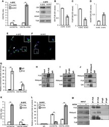
- Fig. 7 Human SPZ mimic mouse SPZ. qRT-PCR detection of circCNOT6L and CNOT6L-mRNA expression levels ( A ); immunoblots and quantification of CB1 ( B ), FUS ( C ) and ADAR ( D ) proteins in A- and B-SPZ fractions from normozoospermic volunteers ( n = 5 different samples in triplicate). Immunofluorescence analysis of FUS protein in A- and B-SPZ ( E and F ). White empty arrowheads and white full arrowheads represent FUS localization (FITC-green) in sperm head and tail, respectively. Nuclei were labeled with DAPI (blue). Scale bar: 20 uM. qRT-PCR detection of circCNOT6L and CNOT6L-mRNA expression levels ( G ) in B-SPZ fractions from normozoospermic volunteers ( n = 5) in vitro treated with vehicle (CTRL) or ACEA 1 uM ( n = 5 different samples for each experimental group in triplicate). In ( A ) and ( G ), the data are reported as mean value of nfe +- SEM, using Gapdh as endogenous control. In ( B ), ( C ) and ( D ) CB1, FUS and ADAR amount was quantified by densitometry analysis, normalized against Tubulin (TUB) signals, expressed as fc of OD values and reported as mean value +- SEM. ** P < 0.01. The enrichment levels of circCNOT6L and CNOT6L-mRNA in the products of RIP assay (FUS-IP compared with IgG-IP) in B-SPZ alone ( H ) and after in vitro ACEA treatment ( L ) by qRT-PCR. Data are reported as mean +- SEM from three independent experiments. ** P < 0.01. Western blot analysis of FUS, QKI and RNApol2 in the products of IP in B-SPZ ( I , J and K ) using FUS, QKI and RNApol2 anti