 Explore
 Validate
 Learn
Learn Western blot
 Immunocytochemistry
Antibody data
- Antibody Data
- Antigen structure
- References [1]
- Comments [0]
- Validations
- Western blot [2]
- Immunoprecipitation [2]
- Immunohistochemistry [7]
Submit
Validation data
Reference
Comment
Report error
- Product number
- NBP2-16258 - Provider product page

- Provider
- Novus Biologicals
- Product name
- Rabbit Polyclonal E-Cadherin Antibody
- Antibody type
- Polyclonal
- Description
- Immunogen affinity purified.
- Reactivity
- Human, Mouse, Rat, Zebrafish
- Host
- Rabbit
- Isotype
- IgG
- Vial size
- 0.1 ml
- Storage
- Aliquot and store at -20C or -80C. Avoid freeze-thaw cycles.
Submitted references Age-Dependent Translocation of Gold Nanoparticles across the Air-Blood Barrier.
Tsuda A, Donaghey TC, Konduru NV, Pyrgiotakis G, Van Winkle LS, Zhang Z, Edwards P, Bustamante JM, Brain JD, Demokritou P
ACS nano 2019 Sep 24;13(9):10095-10102
ACS nano 2019 Sep 24;13(9):10095-10102
No comments: Submit comment
Supportive validation
- Submitted by
- Novus Biologicals (provider)
- Main image

- Experimental details
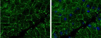
- Western Blot: E-Cadherin Antibody [NBP2-16258] - Non-transfected (-) and transfected (+) MCF-7 whole cell extracts (30 ug) were separated by 5% SDS-PAGE, and the membrane was blotted with E-Cadherin antibody.

- Submitted by
- Novus Biologicals (provider)
- Main image

- Experimental details
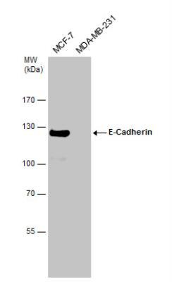
- Western Blot: E-Cadherin Antibody [NBP2-16258] - Various whole cell extracts (30 ug) were separated by 7.5% SDS-PAGE, and the membrane was blotted with E-Cadherin antibody diluted at 1:2000.
Supportive validation
- Submitted by
- Novus Biologicals (provider)
- Main image

- Experimental details
- Immunoprecipitation: E-Cadherin Antibody [NBP2-16258] - MCF-7 whole cell extract A. Control with 3 ug of preimmune Rabbit IgG B. Immunoprecipitation of E-cadherin protein by 3 ug E-cadherin antibody 5 % SDS-PAGE The immunoprecipitated E-cadherin protein was detected by E-cadherin antibody diluted at 1:500. [EasyBlot anti-rabbit IgG was used as a secondary reagent]

- Submitted by
- Novus Biologicals (provider)
- Main image

- Experimental details
- Immunoprecipitation: E-Cadherin Antibody [NBP2-16258] - Immunoprecipitation of E-Cadherin protein from MCF-7 whole cell extracts using 5 ug of E-Cadherin antibody. Western blot analysis was performed using E-Cadherin antibody. EasyBlot anti-Rabbit IgG was used as a secondary reagent.

Supportive validation
- Submitted by
- Novus Biologicals (provider)
- Main image

- Experimental details
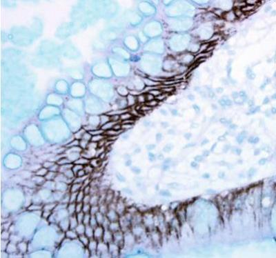
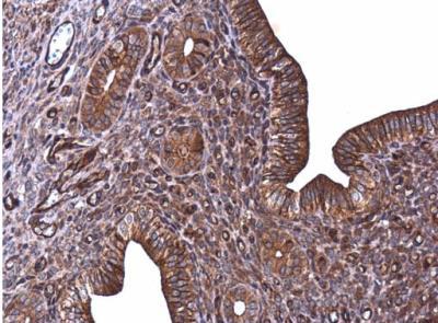
- Immunohistochemistry-Paraffin: E-Cadherin Antibody [NBP2-16258] - Analysis of paraffin-embedded human ulcerative colitis tissue using E-Cadherin antibody.
- Submitted by
- Novus Biologicals (provider)
- Main image

- Experimental details
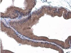
- Immunohistochemistry-Paraffin: E-Cadherin Antibody [NBP2-16258] - Paraffin-embedded rat intestine. E-Cadherin antibody diluted at 1:500.

- Submitted by
- Novus Biologicals (provider)
- Main image

- Experimental details
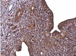
- Immunohistochemistry-Paraffin: E-Cadherin Antibody [NBP2-16258] - Paraffin-embedded breast cancer. E-cadherin antibody dilution: 1:500.

- Submitted by
- Novus Biologicals (provider)
- Main image

- Experimental details
- Immunohistochemistry-Paraffin: E-Cadherin Antibody [NBP2-16258] - Paraffin-embedded mouse pancreas. E-Cadherin antibody diluted at 1:400.
- Submitted by
- Novus Biologicals (provider)
- Main image
- Experimental details
- Immunohistochemistry-Paraffin: E-Cadherin Antibody [NBP2-16258] - Paraffin-embedded mouse cervix. E-Cadherin antibody diluted at 1:500.
- Submitted by
- Novus Biologicals (provider)
- Main image
- Experimental details
- Immunohistochemistry-Paraffin: E-Cadherin Antibody [NBP2-16258] - Paraffin-embedded rat prostate. E-Cadherin antibody diluted at 1:500.

- Submitted by
- Novus Biologicals (provider)
- Main image
- Experimental details
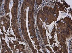
- Immunohistochemistry-Paraffin: E-Cadherin Antibody [NBP2-16258] - Paraffin-embedded rat duodenum. E-Cadherin antibody diluted at 1:500.
