Explore
 Validate
 Learn
Learn Western blot
Antibody data
- Antibody Data
- Antigen structure
- References [1]
- Comments [0]
- Validations
- Western blot [1]
- Immunohistochemistry [1]
- Other assay [1]
Submit
Validation data
Reference
Comment
Report error
- Product number
- MA1-06300 - Provider product page

- Provider
- Invitrogen Antibodies
- Product name
- E-cadherin Monoclonal Antibody (MB2)
- Antibody type
- Monoclonal
- Antigen
- Other
- Description
- MA1-06300 detects E-cadherin in human samples.
- Antibody clone number
- MB2
- Concentration
- 1 mg/mL
Submitted references Effects of DSPP and MMP20 Silencing on Adhesion, Metastasis, Angiogenesis, and Epithelial-Mesenchymal Transition Proteins in Oral Squamous Cell Carcinoma Cells.
Aseervatham J, Ogbureke KUE
International journal of molecular sciences 2020 Jul 2;21(13)
International journal of molecular sciences 2020 Jul 2;21(13)
No comments: Submit comment
Supportive validation
- Submitted by
- Invitrogen Antibodies (provider)
- Main image
- Experimental details
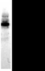
- Western blot analysis of CD324/E Cadherin in MCF-7 cellular extract using CD324/E Cadherin monoclonal antibody (Product # MA1-06300). Moderate reactivity with the 120 kDa full length protein and strong reactivity with the 80 kDa extracellular part of E-Cadherin

Supportive validation
- Submitted by
- Invitrogen Antibodies (provider)
- Main image
- Experimental details
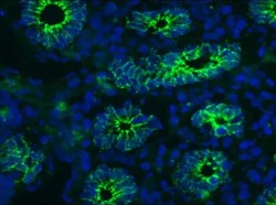
- Immunohistochemistry on frozen sections of small intestine stained with CD324/E Cadherin monoclonal antibody (Product # MA1-06300). Positive staining of the cell-cell adhesion molecules between the epithelial cells of the crypts.

Supportive validation
- Submitted by
- Invitrogen Antibodies (provider)
- Main image

- Experimental details
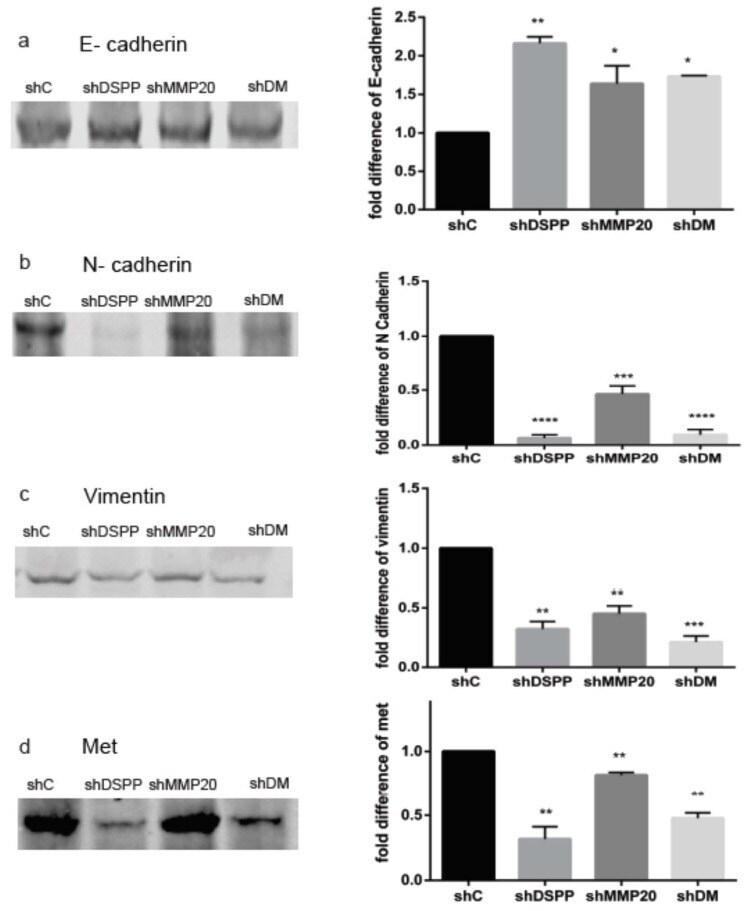
- Figure 4 WB analysis of proteins associated with epithelial-mesenchymal transition. ( a ) Increase in level of E-cadherin (2-fold in shDSPP, 1.6-fold in shMMP20, and 1.8-fold in shDM ( p < 0.01, p < 0.05, p < 0.05, respectively) when compared to the control. ( b ) Decreased N-cadherin in shDSPP (93%), shMMP20 (54%), and shDM (84%) were significant ( p < 0.05) when compared to the control. ( c ) Decreased vimentin in shDSPP (73%), shMMP20 (55%), and shDM (79%) were significant ( p < 0.01, 0.001) when compared to the control. ( d ) Downregulation of met in shDSPP (80%), shMMP20 (30%), and shDM (47%) were significant ( p < 0.01) when compared to the control. ( e ) Downregulation of src ( Figure 4 e) in shDSPP (60%), shMMP20 (75%), and shDM (85%) were significant ( p < 0.01) when compared to the control ( f ) Downregulation of snail in shDSPP (46%), shMMP20 (60%), and shDM (74%) were significant ( p < 0.05) when compared to the control. ( g ) Downregulation of twist in shDSPP (44%), shMMP20 (50%), and shDM (70%) were significant ( p < 0.001) when compared to the control. Beta actin was used as the internal control. Values are given as mean +- SE for 3 independent experiments. shC = Scrambled Control; shDSPP = DSPP Silenced OSC2 cells; shMMP20 = MMP20 Silenced OSC2 cells; DM = Combined DSPP-MMP20 Silenced OSC2 cells; * p < 0.05; ** p < 0.01; *** p < 0.001; **** p < 0.0001.