Explore
 Validate
 Learn
Learn Western blot
 Immunoprecipitation
Antibody data
- Antibody Data
- Antigen structure
- References [4]
- Comments [0]
- Validations
- Western blot [1]
- Immunocytochemistry [1]
Submit
Validation data
Reference
Comment
Report error
- Product number
- GTX11512 - Provider product page

- Provider
- GeneTex
- Proper citation
- GeneTex Cat#GTX11512, RRID:AB_381324
- Product name
- E-Cadherin antibody [DECMA-1]
- Antibody type
- Monoclonal
- Reactivity
- Human, Mouse, Bovine, Canine
- Host
- Rat
Submitted references E-cadherin and LGN align epithelial cell divisions with tissue tension independently of cell shape.
Cell division orientation is coupled to cell-cell adhesion by the E-cadherin/LGN complex.
Vimentin coordinates fibroblast proliferation and keratinocyte differentiation in wound healing via TGF-β-Slug signaling.
Contribution of acidic melanoma cells undergoing epithelial-to-mesenchymal transition to aggressiveness of non-acidic melanoma cells.
Hart KC, Tan J, Siemers KA, Sim JY, Pruitt BL, Nelson WJ, Gloerich M
Proceedings of the National Academy of Sciences of the United States of America 2017 Jul 18;114(29):E5845-E5853
Proceedings of the National Academy of Sciences of the United States of America 2017 Jul 18;114(29):E5845-E5853
Cell division orientation is coupled to cell-cell adhesion by the E-cadherin/LGN complex.
Gloerich M, Bianchini JM, Siemers KA, Cohen DJ, Nelson WJ
Nature communications 2017 Jan 3;8:13996
Nature communications 2017 Jan 3;8:13996
Vimentin coordinates fibroblast proliferation and keratinocyte differentiation in wound healing via TGF-β-Slug signaling.
Cheng F, Shen Y, Mohanasundaram P, Lindström M, Ivaska J, Ny T, Eriksson JE
Proceedings of the National Academy of Sciences of the United States of America 2016 Jul 26;113(30):E4320-7
Proceedings of the National Academy of Sciences of the United States of America 2016 Jul 26;113(30):E4320-7
Contribution of acidic melanoma cells undergoing epithelial-to-mesenchymal transition to aggressiveness of non-acidic melanoma cells.
Peppicelli S, Bianchini F, Torre E, Calorini L
Clinical & experimental metastasis 2014 Apr;31(4):423-33
Clinical & experimental metastasis 2014 Apr;31(4):423-33
No comments: Submit comment
Supportive validation
- Submitted by
- GeneTex (provider)
- Main image
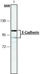
- Experimental details
- MDCK cell extract was separated on SDS-PAGE and probed with Rat Anti-Uvomorulin/E-Cadherin Clone: DECMA-1 (GTX11512). The antibody was developed using Rabbit Anti-Rat IgG-Peroxidase and a chemiluminescent substrate. 1. Antibody dilution 1:250
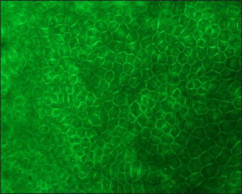
Supportive validation
- Submitted by
- GeneTex (provider)
- Main image

- Experimental details
- MDCK cells were fixed and permeabilized with cold methanol followed by aceton. Fixed cells were stained with Rat Anti-Uvomorulin/E-Cadherin Clone: DECMA-1 (GTX11512) diluted to 1:3,200. The antibody was developed using Rabbit Anti-Rat IgG, FITC-conjμgate.