Explore
 Validate
 Learn
Learn Immunocytochemistry
Antibody data
- Antibody Data
- Antigen structure
- References [3]
- Comments [0]
- Validations
- Immunocytochemistry [1]
- Flow cytometry [1]
- Other assay [3]
Submit
Validation data
Reference
Comment
Report error
- Product number
- 14-9159-80 - Provider product page

- Provider
- Invitrogen Antibodies
- Product name
- Desmoglein 2 Monoclonal Antibody (CSTEM28), eBioscience™
- Antibody type
- Monoclonal
- Antigen
- Other
- Description
- This monoclonal antibody CSTEM28 reacts with human desmoglein 2 (DSG2), a type 1 transmembrane protein belonging to the cadherin family. DSG2 is expressed by epithelial cells, cardiomyocytes, endothelial progenitors, and CD34+CD45dim hematopoietic stem and progenitor cells. This adhesion molecule forms cell-cell adhesion complexes called desmosomes that provide tensile strength to tissues that experience mechanical stress. Other than its role in desmosome formation, DSG2 plays a role in barrier integrity, adhesion, migration, cell apoptosis, proliferation, vasculogenic mimicry, and intracellular signaling. DSG2-expressing endothelial progenitor cells are pro-angiogenic, suggesting a role in vasculature formation. On hematopoietic stem and progenitor cells, DSG2 expression is highest on CD38-CD90+ multi-potent cells. DSG2 expression is progressively decreased as a function of differentiation, with DSG2 expression absent on mature lymphocytes. DSG2 also serves as a receptor for adenoviruses and can be shed by mucosal barriers in the intestine.
- Antibody clone number
- CSTEM28
- Concentration
- 0.5 mg/mL
Submitted references Comparative Analysis of Cell-Cell Contact Abundance in Ovarian Carcinoma Cells Cultured in Two- and Three-Dimensional In Vitro Models.
Chimeric oncolytic Ad5/3 virus replicates and lyses ovarian cancer cells through desmoglein-2 cell entry receptor.
Combination of immunogenic oncolytic adenovirus ONCOS-102 with anti-PD-1 pembrolizumab exhibits synergistic antitumor effect in humanized A2058 melanoma huNOG mouse model.
Kutova OM, Sencha LM, Pospelov AD, Dobrynina OE, Brilkina AA, Cherkasova EI, Balalaeva IV
Biology 2020 Dec 4;9(12)
Biology 2020 Dec 4;9(12)
Chimeric oncolytic Ad5/3 virus replicates and lyses ovarian cancer cells through desmoglein-2 cell entry receptor.
Kuryk L, Møller AW
Journal of medical virology 2020 Aug;92(8):1309-1315
Journal of medical virology 2020 Aug;92(8):1309-1315
Combination of immunogenic oncolytic adenovirus ONCOS-102 with anti-PD-1 pembrolizumab exhibits synergistic antitumor effect in humanized A2058 melanoma huNOG mouse model.
Kuryk L, Møller AW, Jaderberg M
Oncoimmunology 2019;8(2):e1532763
Oncoimmunology 2019;8(2):e1532763
No comments: Submit comment
Supportive validation
- Submitted by
- Invitrogen Antibodies (provider)
- Main image
- Experimental details
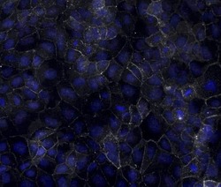
- Immunocytochemistry of fixed and permeabilized 2102EP cells using 10 µg/mL Anti-Human Desmoglein 2 Purified followed by F (ab')2 Anti-Mouse IgG eFluor® 660. Nuclei are stained with DAPI.

Supportive validation
- Submitted by
- Invitrogen Antibodies (provider)
- Main image

- Experimental details
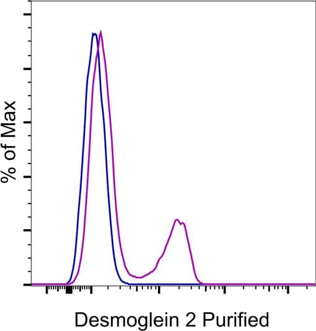
- Staining of a mixture of iPSC and the C2C12 cell line with 0.25 µg of Mouse IgG2b K Isotype Control Purified (Product # 14-4732-82) (blue histogram) or 0.25 µg of Anti-Human Desmoglein 2 Purified (purple histogram) followed by F (ab')2 Anti-Mouse IgG PE (Product # 12-4010-82). Total viable cells were used for analysis.
Supportive validation
- Submitted by
- Invitrogen Antibodies (provider)
- Main image

- Experimental details
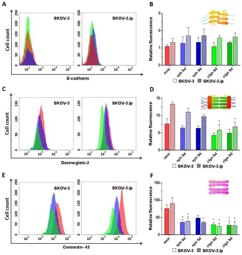
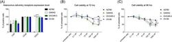
- Figure 3 Expression level of analyzed proteins of adherens junctions (E-cadherin), desmosomes (desmoglein-2) and gap junctions (connexin-43) in SKOV-3 and SKOV-3.ip cells cultured in monolayer and 3D in vitro models. ( A , C , E ) The distributions of SKOV-3 cells (left plot) and SKOV-3.ip cells (right plot) according to fluorescence intensity detected after staining with E-cadherin-specific, desmoglein-2-specific and connexin-43-specific antibodies (red-monolayer culture, blue-spheroids, green-collagen hydrogel); ( B , D , F ) Levels of E-cadherin, desmoglein-2 and connexin-43 in monolayer and 3D models denoted as relative fluorescence values, calculated as a ratio of mean fluorescence intensity of cells stained with specific antibodies to mean fluorescence intensity of cells stained with antibodies of isotypic control. mnlr , monolayer; sph , spheroids; clgn , collagen hydrogel. ""*"" indicates significant difference in RF level from monolayer culture (ANOVA, Holm-Sidak''s multiple comparisons test, p < 0.05).
- Submitted by
- Invitrogen Antibodies (provider)
- Main image
- Experimental details
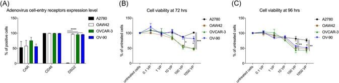
- Figure 1 EOC receptor expression and sensitivity to oncolytic activity to ONCOS-102 treatment. A, Flow cytometry analyses of CAR, CD46, and DSG2 receptor expression on ovarian cancer cells. At least 10 4 events were analyzed for each marker and cell line. Results represent the mean +- SEM of at least two independent experiments. Cell viability at (B) 72 hours or (C) 96 hours after ONCOS-102 treatment in five different concentrations was assessed with the MTS assay. Results are expressed as the mean percent of untreated cells +- SEM. Data represents a pool of two independent experiments run in triplicate. CAR, coxsackie and adenovirus receptor; DSG2, desmoglein-2; EOC, epithelial ovarian cancer
- Submitted by
- Invitrogen Antibodies (provider)
- Main image

- Experimental details
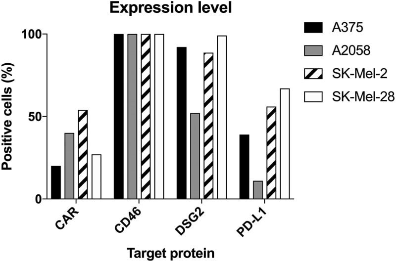
- 10.1080/2162402X.2018.1532763-F0001 Figure 1. Expression of CAR, CD46, Desmoglein-2 and PD-L1 in human melanoma cells measured by flow cytometry (at least 10 4 cells/events were analyzed by flow cytometry in one replicate experiment). Data are expressed as percentage of cells positive for the marker.