Explore
 Validate
 Learn
Learn Western blot
Antibody data
- Antibody Data
- Antigen structure
- References [2]
- Comments [0]
- Validations
- Western blot [5]
- Immunocytochemistry [4]
- Immunohistochemistry [3]
- Flow cytometry [1]
Submit
Validation data
Reference
Comment
Report error
- Product number
- NBP1-69923 - Provider product page

- Provider
- Novus Biologicals
- Product name
- Mouse Monoclonal AKT1 Antibody
- Antibody type
- Monoclonal
- Description
- Protein A purified.
- Reactivity
- Human, Mouse, Rat, Simian
- Host
- Mouse
- Isotype
- IgG
- Vial size
- 0.1 mg
- Concentration
- 1 mg/ml
- Storage
- Store at -20C. Avoid freeze-thaw cycles.
Submitted references A promising natural product, pristimerin, results in cytotoxicity against breast cancer stem cells in vitro and xenografts in vivo through apoptosis and an incomplete autopaghy in breast cancer.
Cytosolic 5'-Nucleotidase II Silencing in a Human Lung Carcinoma Cell Line Opposes Cancer Phenotype with a Concomitant Increase in p53 Phosphorylation.
Cevatemre B, Erkısa M, Aztopal N, Karakas D, Alper P, Tsimplouli C, Sereti E, Dimas K, Armutak EII, Gurevin EG, Uvez A, Mori M, Berardozzi S, Ingallina C, D'Acquarica I, Botta B, Ozpolat B, Ulukaya E
Pharmacological research 2018 Mar;129:500-514
Pharmacological research 2018 Mar;129:500-514
Cytosolic 5'-Nucleotidase II Silencing in a Human Lung Carcinoma Cell Line Opposes Cancer Phenotype with a Concomitant Increase in p53 Phosphorylation.
Pesi R, Petrotto E, Colombaioni L, Allegrini S, Garcia-Gil M, Camici M, Jordheim LP, Tozzi MG
International journal of molecular sciences 2018 Jul 20;19(7)
International journal of molecular sciences 2018 Jul 20;19(7)
No comments: Submit comment
Supportive validation
- Submitted by
- Novus Biologicals (provider)
- Main image
- Experimental details
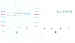
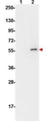
- Western Blot: AKT1 [p Ser473] Antibody (17F6.B11) [NBP1-69923] - A Lane 1) PDGF stimulated NIH 3T3 cells 10 ul Lane 2) NIH 3T3 cells 10 ul Lane 3) Hela whole cell lysate 10 ul (weak signal) B Lane 4) GST negative control protein 100 ng Lane 5) GST negative control protein 25 ng Lane 6) AKT 1 recombinant protein 100 ng Lane 7) AKT 1 recombinant protein 25 ng Block: 5% BSA overnight at 4C. Primary antibody: monoclonal anti AKT antibody, lot no. 27843 used at 1:1000 for overnight at 4C. Secondary antibody: HRP Conjugated goat anti mouse lot 20121 1:25K for 45 min at RT. Detection : TMB Peroxidase substrate for 20 minutes, rinsed with deionized water, dried and scanned on conventional flatbed scanner

- Submitted by
- Novus Biologicals (provider)
- Main image

- Experimental details
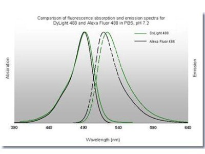
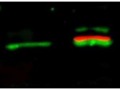
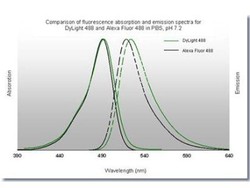
- Western Blot: AKT1 [p Ser473] Antibody (17F6.B11) [NBP1-69923] - Lane 1: unstimulated NIH/3T3 lysates contain inactive unphosphorylated AKT1 (green band). Lane 2: PDGF stimulated NIH/3T3 lysate contains both inactive (green band) and activated phosphorylated Akt1 (red band). Load: 10 ug per lane. Primary antibody: rabbit anti-AKT (pan) and mouse anti-AKT1 pS473 specific antibodies at 1:400 for overnight at 4C. Secondary antibody: DyLight 549 conjugated anti-rabbit IgG (green) and DyLight 649 conjugated anti-mouse IgG (red) secondary antibodies at 1:10,000 for 45 min at RT. Block: 5% BLOTTO overnight at 4C.
- Submitted by
- Novus Biologicals (provider)
- Main image

- Experimental details
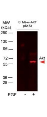
- Western Blot: AKT1 [p Ser473] Antibody (17F6.B11) [NBP1-69923] - Lane 1: A431 cells. Lane 2: A431 cells stimulated for 15 min with EGF. Load: 35 ug per lane. AKT1 phospho Ser473 antibody at 1:400 overnight at 4C. Secondary antibody: DyLight649 Conjugated Anti-AKT pS473 antibody.
- Submitted by
- Novus Biologicals (provider)
- Main image
- Experimental details
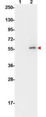
- Western Blot: AKT1 [p Ser473] Antibody (17F6.B11) [NBP1-69923] - Lane 1: non-phosphorylated AKT in untreated cells. Lane 2: phosphorylated AKT (indicated by arrowhead at 56 kDa) on PDGF stimulated NIH/3T3 cell lysates. Load: 10 ug per lane. Primary antibody: AKT phospho Ser473 antibody at 1:10,000 in TBS with 0.05% Tween-20 with 1% BSA, for 1 h at 4 C. Secondary antibody: HRP conjugated Gt-a-Mouse IgG was used at a 1:20,000 dilution for 1 h at 4C with FemtoMax enhanced chemiluminescent reagent.
- Submitted by
- Novus Biologicals (provider)
- Main image

- Experimental details
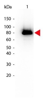
- Western Blot: AKT1 [p Ser473] Antibody (17F6.B11) [NBP1-69923] - Analysis using the Biotin conjugate of NBP1-69923. Detection of Lane 1: GST tagged AKT1 active recombinant protein. Load: 25 ng per lane. Akt phospho S473 Biotin Conjugated antibody at 1:1,000 for overnight at 4C. Secondary antibody: HRP Streptavidin.
Supportive validation
- Submitted by
- Novus Biologicals (provider)
- Main image
- Experimental details
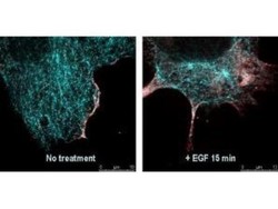
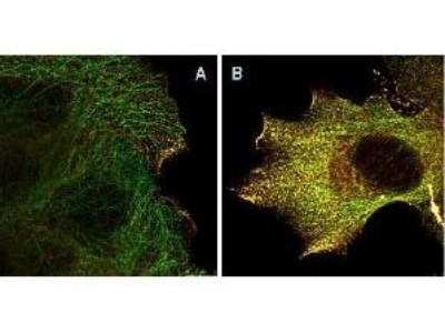
- Immunocytochemistry/Immunofluorescence: AKT1 [p Ser473] Antibody (17F6.B11) [NBP1-69923] - Tissue: EGF treated A431 cells. Fixation: 0.5% PFA. Antigen retrieval: EGF 15 min. Primary antibody: AKT pS473 antibody at 10 ug/mL for 1 h at RT. Secondary antibody: DyLight 488 Goat anti-Rabbit IgG, MAb anti-AKT pS473, atto-647N anti-Mouse IgG (Active Motif). at 1:10,000 for 45 min at RT. Localization: AKT pS473 is nuclear and occasionally cytoplasmic. Staining: AKT pS473 as red signal with tubulin (cyan).

- Submitted by
- Novus Biologicals (provider)
- Main image

- Experimental details
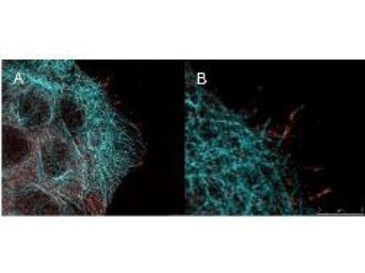
- Immunocytochemistry/Immunofluorescence: AKT1 [p Ser473] Antibody (17F6.B11) [NBP1-69923] - Analysis of A431 cells. The merge images (A) and at high magnification (B) show phosphorylated AKT colocalized with the distal microtubules. Fixation: 4% paraformaldehyde for 5 min and after washes blocked with 10% NGS/0.2% Triton X-100 for 30 min. Antigen retrieval: serum deprivation for 12 h. Primary antibody: AKT pS473 antibody at 10 ug/mL and a-tubulin (cyan) at 1.4 ug/mL for 1 h at RT. Secondary antibody: Atto 647N anti-Mouse IgG (ATTO TEC GmbH), and DyLight488 anti-Rabbit IgG were used at 1.0 ug/mL for 1h at RT for indirect detection. Localization: AKT pS473 is in the cytoplasm and also organized at the periphery of the cell. Staining: AKT pS473 as red signal with bis-benzimide (blue) nuclear counterstain.
- Submitted by
- Novus Biologicals (provider)
- Main image

- Experimental details
- Immunocytochemistry/Immunofluorescence
- Submitted by
- Novus Biologicals (provider)
- Main image

- Experimental details
- Immunocytochemistry/Immunofluorescence
Supportive validation
- Submitted by
- Novus Biologicals (provider)
- Main image

- Experimental details
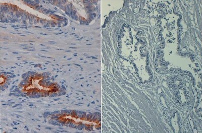
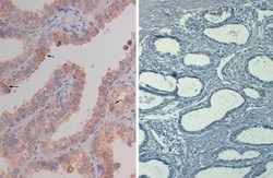
- Immunohistochemistry-Paraffin: AKT1 [p Ser473] Antibody (17F6.B11) [NBP1-69923] - Analysis of FFPE prostate tissue using the Biotin conjugate of AKT1 phospho Ser473 antibody (left panel). Negative control (right panel). Antigen retrieval with heat and pressure in citrate buffer pH 6.2. AKT1 phospho Ser473 antibody at 20 ug/mL. Secondary antibody Streptavidin-HRP at 10 ug/mL. Hematoxylin nuclear counterstain (purple).
- Submitted by
- Novus Biologicals (provider)
- Main image
- Experimental details
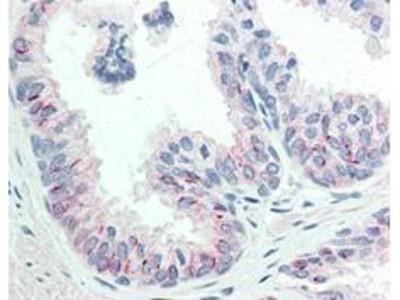
- Immunohistochemistry-Paraffin: AKT1 [p Ser473] Antibody (17F6.B11) [NBP1-69923] - Analysis of Biotin conjugate of NBP1-69923. 20 ug/mL for 1 h at RT Secondary antibody: Streptavidin Conj. HRP 10 ug/ml Localization: nuclear and occasionally cytoplasmic Staining: antibody as precipitated red signal with a hematoxylin purple nuclear count

- Submitted by
- Novus Biologicals (provider)
- Main image
- Experimental details
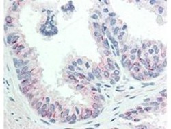
- Immunohistochemistry-Paraffin: AKT1 [p Ser473] Antibody (17F6.B11) [NBP1-69923] - Human FFPE prostate tissue. AKT phospho Ser473 antibody at 20 ug/mL for 1 h at RT. Secondary antibody: Dako's Techmate streptavidin-biotin reagents at 1:10,000 for 45 min at RT. AKT phospho S473 is nuclear and occasionally cytoplasmic. AKT phospho S473 as precipitated red signal with hematoxylin purple nuclear counterstain.
Supportive validation
- Submitted by
- Novus Biologicals (provider)
- Main image
- Experimental details
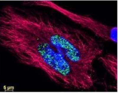
- Flow Cytometry: AKT1 [p Ser473] Antibody (17F6.B11) [NBP1-69923] - Analysis using the DyLight 488 conjugate of AKT1 phospho Ser473 antibody. Image shows anti-histone detection using a DyLight 488 conjugate (green). Anti-Tubulin was detected using a DyLight 549 conjugate (red). Nuclei were counter-stained using DAPI (blue).