Explore
 Validate
 Learn
Learn Flow cytometry
Antibody data
- Antibody Data
- Antigen structure
- References [4]
- Comments [0]
- Validations
- Flow cytometry [1]
- Other assay [4]
Submit
Validation data
Reference
Comment
Report error
- Product number
- 25-9715-42 - Provider product page

- Provider
- Invitrogen Antibodies
- Product name
- Phospho-AKT1 (Ser473) Monoclonal Antibody (SDRNR), PE-Cyanine7, eBioscience™
- Antibody type
- Monoclonal
- Antigen
- Other
- Description
- Description: This SDRNR monoclonal antibody recognizes human and mouse AKT (also known as Protein Kinase B (PKB)) when phosphorylated on S473. AKT is a serine/threonine protein kinase that plays a key role in multiple cellular processes including metabolism, proliferation, apoptosis/survival, and migration. There are three homologous isoforms of AKT: AKT1, AKT2, and AKT3. AKT is activated by binding of its pleckstrin homology (PH) domain to membrane phospholipids and by phosphorylation. Phosphorylation of AKT at T308 by PDK1 and at S473 is required for full activation of this kinase. AKT promotes cell survival by inhibiting apoptosis via phosphorylation and inactivation of several targets including Bad, Foxo1, c-Raf, and caspase-9. Deregulation of AKT has been implicated as a major contributing factor in many types of cancer. AKT is negatively regulated by the phosphatase PTEN as well as by the chemical inhibitor LY294002. Specificity of this SDRNR clone was determined by ELISA, flow cytometry, and western blotting.
- Antibody clone number
- SDRNR
- Concentration
- 5 µL/Test
Submitted references Combinatorial immunotherapy of N-803 (IL-15 superagonist) and dinutuximab with ex vivo expanded natural killer cells significantly enhances in vitro cytotoxicity against GD2(+) pediatric solid tumors and in vivo survival of xenografted immunodeficient NSG mice.
Novel cytokine-antibody fusion protein, N-820, to enhance the functions of ex vivo expanded natural killer cells against Burkitt lymphoma.
IL-15 negatively regulates curdlan-induced IL-23 production by human monocyte-derived dendritic cells and subsequent Th17 response.
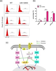
MicroRNA-126 deficiency enhanced the activation and function of CD4(+) T cells by elevating IRS-1 pathway.
Chu Y, Nayyar G, Jiang S, Rosenblum JM, Soon-Shiong P, Safrit JT, Lee DA, Cairo MS
Journal for immunotherapy of cancer 2021 Jul;9(7)
Journal for immunotherapy of cancer 2021 Jul;9(7)
Novel cytokine-antibody fusion protein, N-820, to enhance the functions of ex vivo expanded natural killer cells against Burkitt lymphoma.
Chu Y, Nayyar G, Kham Su N, Rosenblum JM, Soon-Shiong P, Lee J, Safrit JT, Barth M, Lee D, Cairo MS
Journal for immunotherapy of cancer 2020 Oct;8(2)
Journal for immunotherapy of cancer 2020 Oct;8(2)
IL-15 negatively regulates curdlan-induced IL-23 production by human monocyte-derived dendritic cells and subsequent Th17 response.
Eken A, Okus Z, Erdem S, Azizoglu ZB, Haliloglu Y, Bicer A, Gur TN, Yilmaz E, Karakukcu M, Altuntas HD, Canatan H
Northern clinics of Istanbul 2019;6(4):379-387
Northern clinics of Istanbul 2019;6(4):379-387
MicroRNA-126 deficiency enhanced the activation and function of CD4(+) T cells by elevating IRS-1 pathway.
Chu F, Hu Y, Zhou Y, Guo M, Lu J, Zheng W, Xu H, Zhao J, Xu L
Clinical and experimental immunology 2018 Feb;191(2):166-179
Clinical and experimental immunology 2018 Feb;191(2):166-179
No comments: Submit comment
Supportive validation
- Submitted by
- Invitrogen Antibodies (provider)
- Main image

- Experimental details
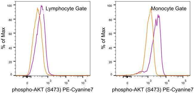
- Normal human peripheral blood cells were unstimulated (orange histogram) or stimulated with Lipopolysaccharide (LPS) Solution (500X) (Product # 00-4976-03) (purple histogram), then intracellularly stained with Anti-Human/Mouse phospho-AKT (S473) PE-Cyanine7 using the Intracellular Fixation and Permeabilization Buffer Set (Product # 88-8824-00) and protocol. Cell in the lymphocyte (left) and monocyte (right) gates were used for analysis.
Supportive validation
- Submitted by
- Invitrogen Antibodies (provider)
- Main image
- Experimental details
- NULL

- Submitted by
- Invitrogen Antibodies (provider)
- Main image
- Experimental details
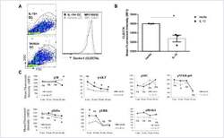
- FIGURE 3 IL-15 exposure downregulates surface Dectin-1 receptor expression and impacts signalling in DCs (A) After five days of culture in with IL-4/GM-CSF or IL-4/GM-CSF/IL-15 DCs were surface stained for Clec7 (Dectin-1) and a representative flow graph and (B) quantification of mean fluorescent intensity (MFI) as bar graph are presented. The experiment was run in triplicate wells and a representative result from a single donor was shown, the experiment was repeated with four different donors. (C) DCs from ""A"" were stimulated with curdlan (50 ug/ml) and phosphorylation of p38, AKT, SRC, NFKB p65, IkBa, ERK1/2 and IRAK4 was measured by phospho-flow assay. A representative result from PBMCs of a single donor was charted. *p

- Submitted by
- Invitrogen Antibodies (provider)
- Main image
- Experimental details
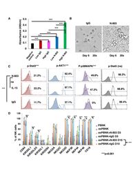
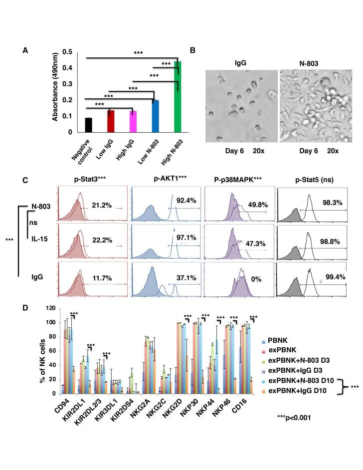
- Figure 1 N-803 increased the viability and proliferation of exPBNK with enhanced p-Stat3, p-Stat5, pAKT, p-p38MAPK and NK activating receptors. PBMNCs were stimulated with irradiated genetically modified K562-mbIL21-41BBL cells for 2-3 weeks. (A) Purified exPBNK cells were cultured in complete medium with 0.35 ng/mL (low) or 3.5 ng/mL (high) N-803 or molar equivalent dose of IgG for 3 days. NK viability and proliferation were monitored by MTS assays. The amount of 490 nm absorbance is directly proportional to the number of living exPBNK cells in the culture. The exPBNK cells with N-803 at 0.35 ng/mL or 3.5 ng/mL have significantly higher viability as compared with IgG or medium controls (p
- Submitted by
- Invitrogen Antibodies (provider)
- Main image

- Experimental details
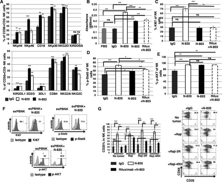
- Figure 1 N-820 enhanced the expression of NK activating receptors and proliferation of exPBNK with enhanced Ki67, p-Stat5, and CD25 levels. The percentages of NK activating and inhibitory receptors on exPBNK surface were monitored by flow cytometry analysis at day 3 (A). The NK proliferation was monitored by CellTiter 96 AQueous One solution cell proliferation assay (Promega) according to the manufacturer's instruction (B). Intracellular Ki67 (C), Phosphorylated STAT5 (p-STAT5) (D) and phosphorylated Akt (p-AKT) (E) were monitored by flow cytometry analysis at day 3. Ki67 (p