Explore
 Validate
 Learn
Learn Western blot
Antibody data
- Antibody Data
- Antigen structure
- References [4]
- Comments [0]
- Validations
- Western blot [2]
- Immunocytochemistry [1]
Submit
Validation data
Reference
Comment
Report error
- Product number
- 14-6583-80 - Provider product page

- Provider
- Invitrogen Antibodies
- Product name
- alpha-Fetoprotein Monoclonal Antibody (AFP3), eBioscience™
- Antibody type
- Monoclonal
- Antigen
- Other
- Description
- Description: This AFP3 monoclonal antibody reacts with human alpha-fetoprotein (AFP). This 70-kDa secretory protein is a member of the albumin gene family. Synthesized by the yolk sac and fetal liver during embryogenesis, AFP protein levels are highest in fetal serum. After birth, serum AFP levels decrease dramatically. In fact, AFP is nearly undetectable in normal adult serum. However, hepatocellular carcinoma and germ cell teratoblastoma, as well as liver regeneration, viral hepatitis, and cirrhosis, leads to elevated AFP serum levels in adults. As such, detection of this protein is frequently used as a diagnostic marker for these conditions.
- Antibody clone number
- AFP3
- Concentration
- 0.5 mg/mL
Submitted references Ectopic hTERT expression facilitates reprograming of fibroblasts derived from patients with Werner syndrome as a WS cellular model.
Expression of intercellular adhesion molecule 1 by hepatocellular carcinoma stem cells and circulating tumor cells.
Molecular mechanisms of alpha-fetoprotein gene expression.
Generation of monoclonal antibodies to alpha-fetoprotein and application in solid-phase enzyme immunoassay.
Wang S, Liu Z, Ye Y, Li B, Liu T, Zhang W, Liu GH, Zhang YA, Qu J, Xu D, Chen Z
Cell death & disease 2018 Sep 11;9(9):923
Cell death & disease 2018 Sep 11;9(9):923
Expression of intercellular adhesion molecule 1 by hepatocellular carcinoma stem cells and circulating tumor cells.
Liu S, Li N, Yu X, Xiao X, Cheng K, Hu J, Wang J, Zhang D, Cheng S, Liu S
Gastroenterology 2013 May;144(5):1031-1041.e10
Gastroenterology 2013 May;144(5):1031-1041.e10
Molecular mechanisms of alpha-fetoprotein gene expression.
Lazarevich NL
Biochemistry. Biokhimiia 2000 Jan;65(1):117-33
Biochemistry. Biokhimiia 2000 Jan;65(1):117-33
Generation of monoclonal antibodies to alpha-fetoprotein and application in solid-phase enzyme immunoassay.
Kuo CY, Fu J, Yeh MY, Su SL, Lee CY
Biotechnology and applied biochemistry 1989 Feb;11(1):96-104
Biotechnology and applied biochemistry 1989 Feb;11(1):96-104
No comments: Submit comment
Supportive validation
- Submitted by
- Invitrogen Antibodies (provider)
- Main image

- Experimental details
- Immunoblotting of reduced HepG2 cell lysate with 5 µg/mL of Anti-Human alpha-Fetoprotein Purified.Bands were visualized using Anti-Mouse IgG HRP.

- Submitted by
- Invitrogen Antibodies (provider)
- Main image
- Experimental details
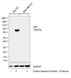
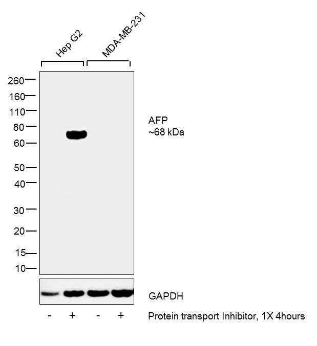
- Western blot was performed using Anti-AFP Monoclonal Antibody (Product # 14-6583-82) and a 68 kDa band corresponding to AFP was observed upon treatment with protein transport inhibitor in Hep G2 cells but not in MDA-MB-231 cells which are reported to be negative. Whole cell extracts (30 µg lysate) of Hep G2 (Lane 1), Hep G2 treated with 1X protein transport inhibitor (Product # 00-4980-03) for 4 hours (Lane 2), MDA-MB-231 (Lane 3) and MDA-MB-231 treated with 1X protein transport inhibitor for 4 hours (Product # 00-4980-03) (Lane 4) were electrophoresed using Novex® NuPAGE® 4-12 % Bis-Tris gel (Product # NP0322BOX). Resolved proteins were then transferred onto a nitrocellulose membrane (Product # IB23001) by iBlot® 2 Dry Blotting System (Product # IB21001). The blot was probed with the primary antibody (5 µg/mL) and detected by chemiluminescence with Goat anti-Mouse IgG (H+L), Superclonal™ Recombinant Secondary Antibody, HRP (Product # A28177, 1:4000 dilution) using the iBright FL 1000 (Product # A32752). Chemiluminescent detection was performed using Novex® ECL Chemiluminescent Substrate Reagent Kit (Product # WP20005).
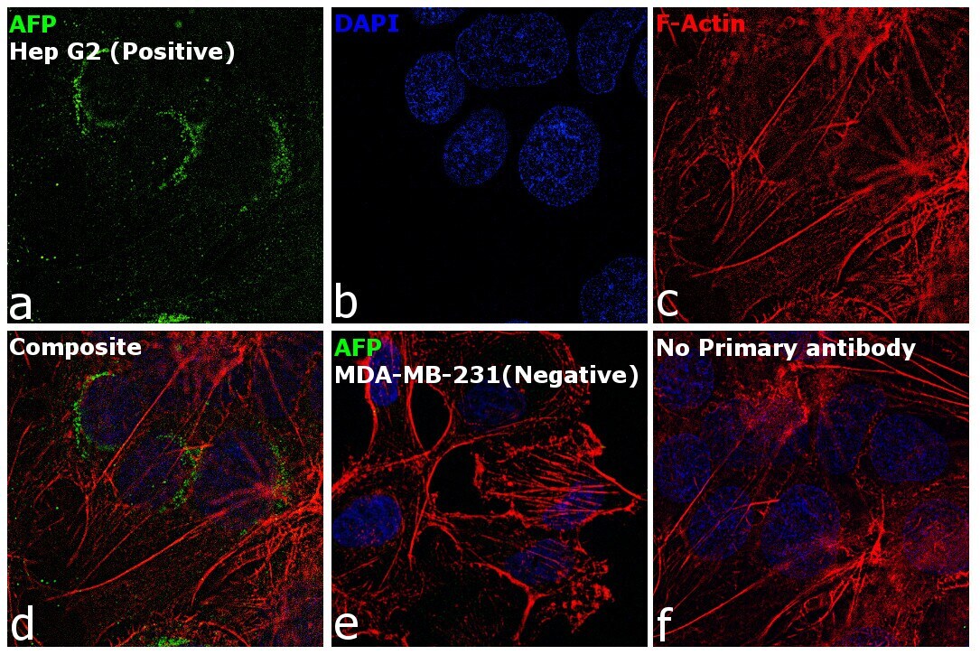
Supportive validation
- Submitted by
- Invitrogen Antibodies (provider)
- Main image
- Experimental details
- Immunofluorescence analysis of AFP was performed using 70% confluent log phase Hep G2 and MDA-MB-231 cells. The cells were fixed with 4% paraformaldehyde for 10 minutes, permeabilized with 0.1% Triton™ X-100 for 15 minutes, and blocked with 2% BSA for 1 hour at room temperature. The cells were labeled with AFP Mouse Monoclonal Antibody (Product # 14-6583-82) at 5 µg/mL in 0.1% BSA and incubated overnight at 4 degree and then labeled with Goat anti-Mouse IgG (H+L), Superclonal™ Recombinant Secondary Antibody, Alexa Fluor 488 (Product # A28175) at a dilution of 1:2000 for 45 minutes at room temperature (Panel a: green). Nuclei (Panel b: blue) were stained with ProLong™ Diamond Antifade Mountant with DAPI (Product # P36962). F-actin (Panel c: red) was stained with Rhodamine Phalloidin (Product # R415, 1:300). Panel d represents the composite image showing cytoplasmic and golgi staining of AFP in Hep G2. Panel e represents the merged image of MDA-MB-231 cells which do not have AFP expression. Panel f represents control cells with no primary antibody to assess background. The images were captured at 60X magnification..