Explore
 Validate
 Learn
Learn Western blot
Antibody data
- Antibody Data
- Antigen structure
- References [1]
- Comments [0]
- Validations
- Western blot [3]
- Immunocytochemistry [2]
- Other assay [1]
Submit
Validation data
Reference
Comment
Report error
- Product number
- 711823 - Provider product page

- Provider
- Invitrogen Antibodies
- Product name
- Ataxin 3 Recombinant Polyclonal Antibody (13HCLC)
- Antibody type
- Polyclonal
- Antigen
- Other
- Description
- This antibody is predicted to react with Monkey, Horse, Dog
- Antibody clone number
- 13HCLC
- Concentration
- 0.5 mg/mL
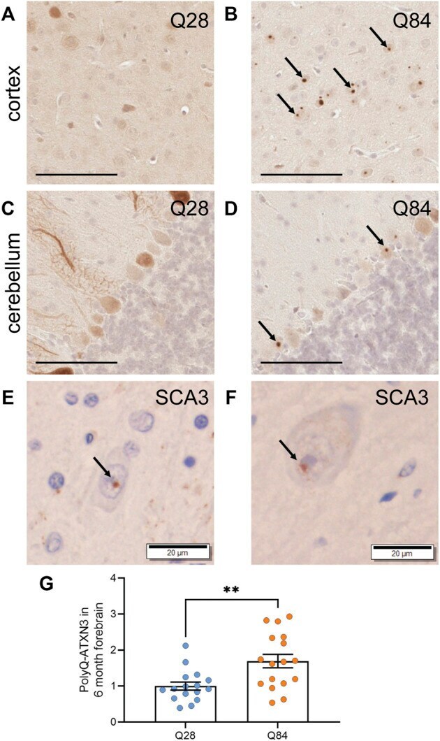
Submitted references Plasma PolyQ-ATXN3 Levels Associate With Cerebellar Degeneration and Behavioral Abnormalities in a New AAV-Based SCA3 Mouse Model.
Jansen-West K, Todd TW, Daughrity LM, Yue M, Tong J, Carlomagno Y, Del Rosso G, Kurti A, Jones CY, Dunmore JA, Castanedes-Casey M, Dickson DW, Wszolek ZK, Fryer JD, Petrucelli L, Prudencio M
Frontiers in cell and developmental biology 2022;10:863089
Frontiers in cell and developmental biology 2022;10:863089
No comments: Submit comment
Supportive validation
- Submitted by
- Invitrogen Antibodies (provider)
- Main image
- Experimental details
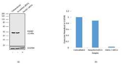
- Knockdown of Ataxin 3 was achieved by transfecting U87MG cells with Ataxin 3 specific siRNA (Silencer® select Product # s230538 and Product # s8794). Western blot analysis (Fig a) was performed using whole cell extract from the Ataxin 3 knock down cells (lane 3), non-specific scrambled siRNA transfected cells (lane 2) and untransfected cells (lane 1). The blots were probed with Anti-Ataxin 3 Recombinant Rabbit Polyclonal Antibody (Product # 711823, 1-3 µg/mL) and Goat anti-Rabbit IgG (H+L) Superclonal™ Secondary Antibody, HRP conjugate (Product # A27036, 0.4 µg/mL, 1:2500 dilution). Densitometric analysis of this Western blot is shown in histogram (Fig b). Loss of signal upon siRNA mediated knock down confirms that antibody is specific to Ataxin 3.

- Submitted by
- Invitrogen Antibodies (provider)
- Main image

- Experimental details
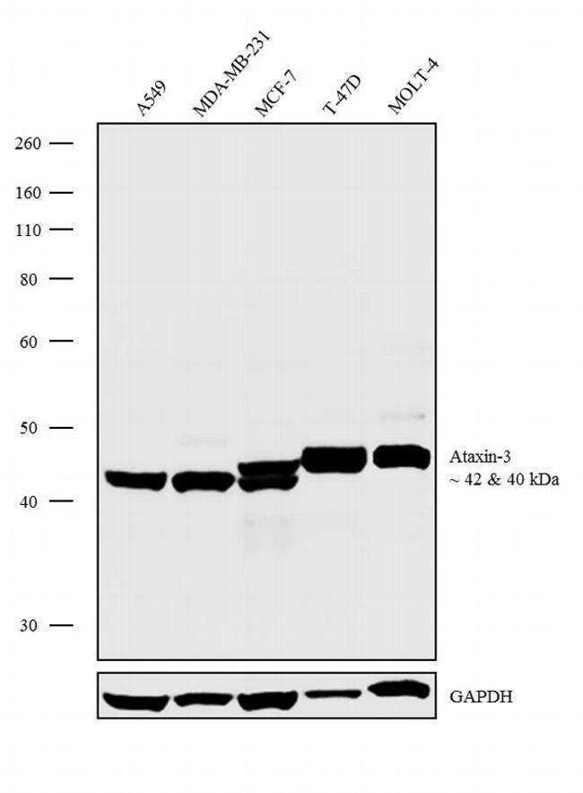
- Western blot analysis was performed on Modified Whole cell extracts (1% SDS) (30 µg lysate) of A549 (Lane 1), MDA-MB-231 (Lane 2), MCF-7 (Lane 3), T-47D (Lane 4) and MOLT-4 (Lane 5). The blots were probed with Anti-Ataxin 3 Recombinant Rabbit Polyclonal Antibody (Product # 711823, 2.5 µg/mL) and detected by chemiluminescence using Goat anti-Rabbit IgG (H+L) Superclonal™ Secondary Antibody, HRP conjugate (Product # A27036, 0.4 µg/mL, 1:4000 dilution). 42 and 40 kDa band corresponding to Ataxin-3 was observed across the cell lines tested. Known quantity of protein samples were electrophoresed using Novex®NuPAGE®4-12% Bis-Tris gel (Product # NP0321BOX), XCell SureLock™ Electrophoresis System (Product # EI0002) and Novex® Sharp Pre-Stained Protein Standard (Product # LC5800). Resolved proteins were then transferred onto a nitrocellulose membrane with iBlot® Dry Blotting System (Product # IB21001). The membrane was probed with the relevant primary and secondary Antibody following blocking with 5% skimmed milk. Chemiluminescent detection was performed using Pierce™ ECL Western blotting Substrate (Product # 32106).
- Submitted by
- Invitrogen Antibodies (provider)
- Main image
- Experimental details
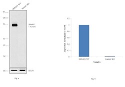
- Western blot analysis (Fig a) of Ataxin 3 was performed on cell extracts (100 µg of lysate) of HEK-293 wild type (Lane 1) and Ataxin 3 knockout (Lane 2). The blot was probed with Anti-Ataxin 3 Recombinant Rabbit Polyclonal Antibody (Product # 711823, 1:400 dilution) and detected by chemiluminescence using Peroxidase AffiniPure Goat anti-Rabbit IgG (H+L) Secondary Antibody, HRP conjugate (Product # 111-035-144, 1:4000 dilution). Densitometric analysis of this Western blot is shown in the histogram (Fig b). Loss of signal upon CRISPR mediated knockout (KO) confirms that antibody is specific to Ataxin 3.

Supportive validation
- Submitted by
- Invitrogen Antibodies (provider)
- Main image
- Experimental details
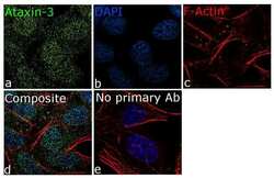
- For immunofluorescence analysis, SH-SY5Y cells were fixed and permeabilized for detection of endogenous Ataxin 3 using Anti-Ataxin 3 Recombinant Rabbit Polyclonal Antibody (Product # 711823, 5 µg/mL) and labeled with Goat anti-Rabbit IgG (H+L) Superclonal™ Secondary Antibody, Alexa Fluor® 488 conjugate (Product # A27034, 1:2000). Panel a) shows representative cells that were stained for detection and localization of Ataxin 3 (green), Panel b) is stained for nuclei (blue) using SlowFade® Gold Antifade Mountant with DAPI (Product # S36938). Panel c) represents cytoskeletal F-actin staining using Rhodamine Phalloidin (Product # R415, 1:300). Panel d) is a composite image of Panels a, b and c clearly demonstrating cytoplasmic and nuclear localization of Ataxin 3. Panel e) represents control cells with no primary antibody to assess background. The images were captured at 60X magnification.

- Submitted by
- Invitrogen Antibodies (provider)
- Main image
- Experimental details
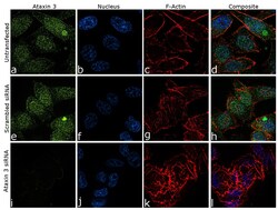
- Knockdown of Ataxin 3 was achieved by transfecting PC-3 cells with specific siRNA (Silencer® select Product # s230538 & s8794). Immunofluorescence analysis was performed on PC-3 cells (untransfected, panel a-d), transfected with Ataxin 3 specific siRNA (panel i-l) or non-specific scrambled siRNA (panels e-h). Cells were fixed, permeabilized, and labelled with Anti-Ataxin 3 Recombinant Rabbit Polyclonal Antibody (Product # 711823, 1:100 dilution), followed by Goat anti-Rabbit IgG (H+L) Superclonal™ Secondary Antibody, Alexa Fluor® 488 conjugate (Product # A27034, 1:2000). Nuclei (blue) were stained using ProLong™ Diamond Antifade Mountant with DAPI (Product # P36962), and Rhodamine Phalloidin (Product # R415, 1:300) was used for cytoskeletal F-actin (red) staining. Significant reduction of signal was observed upon siRNA mediated knockdown (panel i-l) confirming specificity of the antibody to Ataxin 3 (green). The images were captured at 60X magnification.

Supportive validation
- Submitted by
- Invitrogen Antibodies (provider)
- Main image

- Experimental details
- FIGURE 3 PolyQ-expanded ATXN3 forms inclusions in SCA3 mice (A - D) Representative images of IHC staining for ATXN3 in the cortex (A - B) and cerebellum (C - D) of Q28 (A , C) and Q84 (B , D) mice at 6 months of age. Arrows mark inclusions. Scale bars are 100 um (E) IHC staining for ubiquitin in the brain of a SCA3 patient. Arrow marks an inclusion. Scale bar is 20 um. (F) IHC staining for the polyQ antibody clone 1C2 in the brain of a SCA3 patient. Arrow marks an inclusion. Scale bar is 20 um. (G) PolyQ-ATXN3 levels as measured by immunoassay in the forebrain of Q28 and Q84 mice at 6 months of age. Similar results were seen at 3 months; see Supplementary Figure S5 . Error bars are the SEM. ** p