Explore
 Validate
 Learn
Learn Western blot
Antibody data
- Antibody Data
- Antigen structure
- References [3]
- Comments [0]
- Validations
- Western blot [1]
Submit
Validation data
Reference
Comment
Report error
- Product number
- NSB1006 - Provider product page

- Provider
- Novus Biologicals
- Proper citation
- Novus Cat#NSB1006, RRID:AB_10000569
- Product name
- Rabbit Polyclonal PDGF R alpha Antibody
- Antibody type
- Polyclonal
- Description
- Immunogen affinity purified.
- Reactivity
- Human, Mouse
- Host
- Rabbit
- Isotype
- IgG
- Vial size
- 0.1 ml
- Concentration
- 0.5 mg/ml
- Storage
- Store at -80C. Avoid freeze-thaw cycles.
Submitted references The C-terminal tail domain of metavinculin, vinculin's splice variant, severs actin filaments.
Imaging the impact of Nox4 in cycling hypoxia-mediated U87 glioblastoma invasion and infiltration.
Neuroprotective effects of PDGF against oxidative stress and the signaling pathway involved.
Janssen ME, Liu H, Volkmann N, Hanein D
The Journal of cell biology 2012 May 28;197(5):585-93
The Journal of cell biology 2012 May 28;197(5):585-93
Imaging the impact of Nox4 in cycling hypoxia-mediated U87 glioblastoma invasion and infiltration.
Hsieh CH, Chang HT, Shen WC, Shyu WC, Liu RS
Molecular imaging and biology : MIB : the official publication of the Academy of Molecular Imaging 2012 Aug;14(4):489-99
Molecular imaging and biology : MIB : the official publication of the Academy of Molecular Imaging 2012 Aug;14(4):489-99
Neuroprotective effects of PDGF against oxidative stress and the signaling pathway involved.
Zheng L, Ishii Y, Tokunaga A, Hamashima T, Shen J, Zhao QL, Ishizawa S, Fujimori T, Nabeshima Y, Mori H, Kondo T, Sasahara M
Journal of neuroscience research 2010 May 1;88(6):1273-84
Journal of neuroscience research 2010 May 1;88(6):1273-84
No comments: Submit comment
Supportive validation
- Submitted by
- Novus Biologicals (provider)
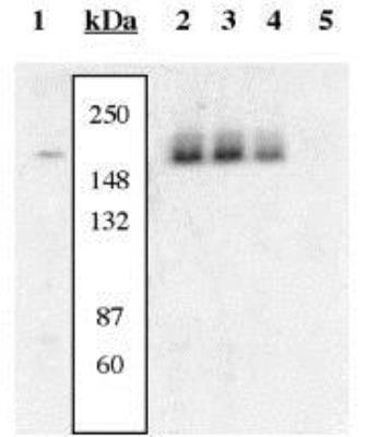
- Main image

- Experimental details
- Western Blot: PDGF Receptor alpha [p Tyr742] Antibody [NSB1006] - Extracts prepared from NIH3T3 cells left unstimulated (1) and stimulated with PDGF (2-5) were resolved by SDS-PAGE on a 10% polyacrylamide gel and transferred to PVDF. Membranes were blocked with a 5% BSA-TBST buffer overnight at 4C, then were incubated with 0.50 ug/ml of NSB1006 for 2hrs at room temp in a 1% BSA-TBST buffer, following prior incubation with: no peptide (1, 2), the non-phosphopeptide corresponding to the immunogen (3), a generic phosphotyrosine containing peptide (4), or, the phosphopeptide immunogen (5). The data show that only the peptide corresponding to PDGFRalpha [pY742] blocks the antibody signal, thereby demonstrating the specificity of the antibody.