 Explore
 Validate
 Learn
LearnMA1-10148
antibody from Invitrogen Antibodies
Targeting: IGF2R
CD222, CI-M6PR, CI-MPR, CIMPR, M6P-R, MPR1, MPR300, MPRI
 Flow cytometry
Antibody data
- Antibody Data
- Antigen structure
- References [1]
- Comments [0]
- Validations
- Flow cytometry [1]
- Other assay [1]
Submit
Validation data
Reference
Comment
Report error
- Product number
- MA1-10148 - Provider product page

- Provider
- Invitrogen Antibodies
- Product name
- IGF2R Monoclonal Antibody (MEM-238), PE
- Antibody type
- Monoclonal
- Antigen
- Recombinant full-length protein
- Description
- This antibody recognizes an epitope between domains 2 and 5 of CD222 (IGF2 receptor), a ubiquitously expressed 250 kDa multifunctional type I transmembrane protein. The majority of CD222 is found in the late endosomal/prelysosomal compartment, 5-10% in the plasma membrane and the truncated (220 kDa) form of CD222 is present in human and bovine serum.
- Reactivity
- Human
- Host
- Mouse
- Conjugate
- Yellow dye
- Isotype
- IgG
- Antibody clone number
- MEM-238
- Vial size
- 100 Tests
- Storage
- 4° C, store in dark, DO NOT FREEZE!
Submitted references Modulation of P2X4/P2X7/Pannexin-1 sensitivity to extracellular ATP via Ivermectin induces a non-apoptotic and inflammatory form of cancer cell death.
Draganov D, Gopalakrishna-Pillai S, Chen YR, Zuckerman N, Moeller S, Wang C, Ann D, Lee PP
Scientific reports 2015 Nov 10;5:16222
Scientific reports 2015 Nov 10;5:16222
No comments: Submit comment
Supportive validation
- Submitted by
- Invitrogen Antibodies (provider)
- Main image
- Experimental details
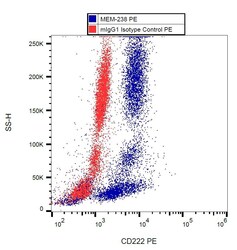
- Flow cytometry analysis (intracellular staining) of human peripheral blood with anti-CD222 (MEM-238) PE Monoclonal antibody (Product # MA1-10148).
- Conjugate
- Yellow dye

Supportive validation
- Submitted by
- Invitrogen Antibodies (provider)
- Main image
- Experimental details
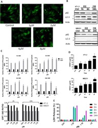
- Figure 6 Ivermectin induces autophagy. ( A ) Ivermectin induces autophagy in breast cancer cells. MDA-MB-231 GFL-LC3 cells were treated with different doses of Ivermectin for 24 h and formation of green fluorescent puncta was evaluated by confocal fluorescence microscopy. ( B ) Western blot confirming the induction of autophagy in both murine and human breast cancer cells, as evidenced by LC3 lipidation and autophagic degradation of p62(SQSTM1). ( C ) Ivermectin up-regulates surface exposure of M6P receptor (top panel) but does not impact the exposure of Calreticulin (CRT) (bottom panel). MDA-MB-231 cells (triplicates) were treated with different doses of Ivermectin for up to 24 h and surface stained with antibody-specific for the human M6P receptor versus isotype control. Similarly, 4T1.2 and MB231 cells were treated with different doses of Ivermectin for 4-24 h, and were stained with an antibody specific for both human and mouse CRT versus isotype control. Mean fluorescence intensity (MFI) values and ratios versus isotype control were calculated after gating on live/membrane-intact cells. ( D ) Ivermectin (32 muM) induces release of HMGB1 from murine and human TNBC cells (triplicates). ( E ) Ivermectin treatment induces release of cytosolic LDH, data are normalized to maximum release (lysis). Asterisk (*) indicates p < 0.05 relative to untreated or Ivermectin alone controls, respectively.
- Conjugate
- Yellow dye
