Explore
 Validate
 Learn
Learn Western blot
Antibody data
- Antibody Data
- Antigen structure
- References [3]
- Comments [0]
- Validations
- Western blot [3]
- Immunoprecipitation [1]
Submit
Validation data
Reference
Comment
Report error
- Product number
- NB100-55249 - Provider product page

- Provider
- Novus Biologicals
- Proper citation
- Novus Cat#NB100-55249, RRID:AB_829110
- Product name
- Rabbit Polyclonal HGS Antibody
- Antibody type
- Polyclonal
- Description
- Immunogen affinity purified.
- Reactivity
- Human, Mouse
- Host
- Rabbit
- Isotype
- IgG
- Vial size
- 0.1 ml
- Concentration
- 0.2 mg/ml
- Storage
- Store at 4C. Do not freeze.
Submitted references The multivesicular body is the major internal site of prion conversion.
CIN85 phosphorylation is essential for EGFR ubiquitination and sorting into multivesicular bodies.
Proteolytic processing of protocadherin proteins requires endocytosis.
Yim YI, Park BC, Yadavalli R, Zhao X, Eisenberg E, Greene LE
Journal of cell science 2015 Apr 1;128(7):1434-43
Journal of cell science 2015 Apr 1;128(7):1434-43
CIN85 phosphorylation is essential for EGFR ubiquitination and sorting into multivesicular bodies.
Schroeder B, Srivatsan S, Shaw A, Billadeau D, McNiven MA
Molecular biology of the cell 2012 Sep;23(18):3602-11
Molecular biology of the cell 2012 Sep;23(18):3602-11
Proteolytic processing of protocadherin proteins requires endocytosis.
Buchanan SM, Schalm SS, Maniatis T
Proceedings of the National Academy of Sciences of the United States of America 2010 Oct 12;107(41):17774-9
Proceedings of the National Academy of Sciences of the United States of America 2010 Oct 12;107(41):17774-9
No comments: Submit comment
Supportive validation
- Submitted by
- Novus Biologicals (provider)
- Main image
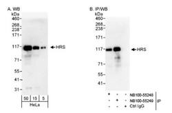
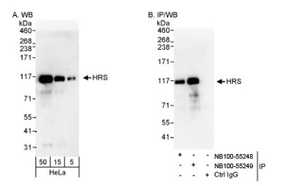
- Experimental details
- Western Blot: HGS Antibody [NB100-55249] - Detection of Human HRS on HeLa whole cell lysate using NB100-55249. HRS was also immunoprecipitated by rabbit anti-HRS antibody Nb100-55248.
- Submitted by
- Novus Biologicals (provider)
- Main image
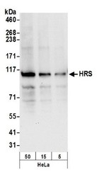
- Experimental details
- Western Blot: HGS Antibody [NB100-55249] - Detection of Human HRS by Western Blot. Samples: Whole cell lysate (50, 15, 5 ug) from HeLa cells prepared using NETN lysis buffer. Antibody: Affinity purified rabbit anti-HRS antibody NB100-55249 used for WB at 0.1 ug/ml. Detection: Chemiluminescence with an exposure time of 10 seconds.

- Submitted by
- Novus Biologicals (provider)
- Main image
- Experimental details
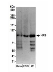
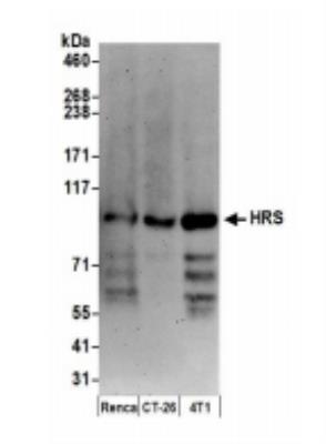
- Western Blot: HGS Antibody [NB100-55249] - Whole cell lysate (15 ug) from Renca, CT26.WT and 4T1 cells prepared using NETN lysis buffer. Antibody: Affinity purified rabbit anti-HRS antibody used for WB at 0.1 ug/ml. Detection: Chemiluminescence with an exposure time of 3 minutes.
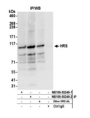
Supportive validation
- Submitted by
- Novus Biologicals (provider)
- Main image

- Experimental details
- Immunoprecipitation: HGS Antibody [NB100-55249] - Detection of human HRS by western blot of immunoprecipitates. Samples: Whole cell lysate (0.5 or 1.0 mg per IP reaction; 20% of IP loaded) from HeLa cells prepared using NETN lysis buffer. Antibodies: Affinity purified rabbit anti-HRS antibody NB100-55249 (lot NB100-55249-2) used for IP at 6 ug per reaction. HRS was also immunoprecipitated by a previous lot of this antibody (NB100-55249-1) and another rabbit anti-HRS antibody. For blotting immunoprecipitated HRS, NB100-55249 was used at 0.4 ug/ml. Detection: Chemiluminescence with an exposure time of 3 seconds.