Explore
 Validate
 Learn
Learn Western blot
 Immunocytochemistry
Antibody data
- Antibody Data
- Antigen structure
- References [0]
- Comments [0]
- Validations
- Western blot [3]
- Immunocytochemistry [1]
Submit
Validation data
Reference
Comment
Report error
- Product number
- MAB72441 - Provider product page

- Provider
- Novus Biologicals
- Product name
- Mouse Monoclonal PKM2 Antibody
- Antibody type
- Monoclonal
- Description
- Protein A or G purified from hybridoma culture supernatant. Detects human, mouse, and rat PKM-2 in Western blot
- Reactivity
- Human, Mouse, Rat
- Host
- Mouse
- Conjugate
- Unconjugated
- Isotype
- IgG
- Vial size
- 100 ug
- Storage
- Use a manual defrost freezer and avoid repeated freeze-thaw cycles. 12 months from date of receipt, -20 to -70 degreesC as supplied. 1 month, 2 to 8 degreesC under sterile conditions after reconstitution. 6 months, -20 to -70 degreesC under sterile conditions after reconstitution.
No comments: Submit comment
Enhanced validation
- Submitted by
- Novus Biologicals (provider)
- Enhanced method
- Genetic validation
- Main image
- Experimental details
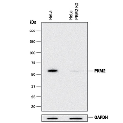
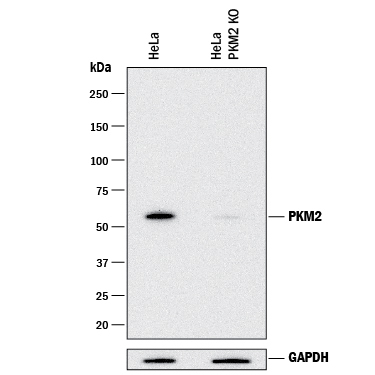
- Western blot shows lysates of HeLa human cervical epithelial carcinoma parental cell line and PKM2 knockout HeLa cell line (KO). PVDF membrane was probed with 0.25 µg/mL of Mouse Anti-Human/Mouse/Rat PKM2 Monoclonal Antibody (Catalog # MAB72441) followed by HRP-conjugated Anti-Mouse IgG Secondary Antibody (Catalog # HAF018). A specific band was detected for PKM2 at approximately 60 kDa (as indicated) in the parental HeLa cell line, but is not detectable in knockout HeLa cell line. GAPDH (Catalog # AF5718) is shown as a loading control. This experiment was conducted under reducing conditions and using Immunoblot Buffer Group 1.
- Submitted by
- Novus Biologicals (provider)
- Main image

- Experimental details
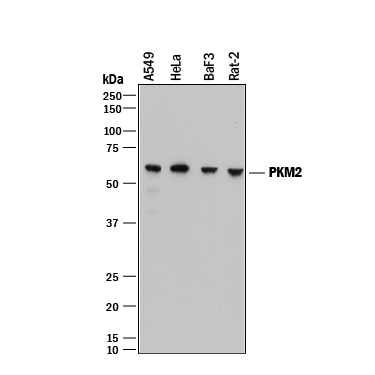
- Detection of Human, Mouse, and Rat PKM2 by Western Blot. Western blot shows lysates of A549 human lung carcinoma cell line, HeLa human cervical epithelial carcinoma cell line, BaF3 mouse pro-B cell line, and Rat-2 rat embryonic fibroblast cell line. PVDF membrane was probed with 0.25 µg/mL of Mouse Anti-Human PKM2 Monoclonal Antibody (Catalog # MAB72441) followed by HRP-conjugated Anti-Mouse IgG Secondary Antibody (Catalog # HAF018). A specific band was detected for PKM2 at approximately 60 kDa (as indicated). This experiment was conducted under reducing conditions and using Immunoblot Buffer Group 1.
- Submitted by
- Novus Biologicals (provider)
- Main image
- Experimental details
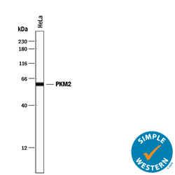
- Detection of Human PKM2 by Simple WesternTM. Simple Western lane view shows lysate of HeLa human cervical epithelial carcinoma cell line, loaded at 0.2 mg/mL. A specific band was detected for PKM2 at approximately 61 kDa (as indicated) using 2.5 µg/mL of Mouse Anti-Human/Mouse/Rat PKM2 Monoclonal Antibody (Catalog # MAB72441) . This experiment was conducted under reducing conditions and using the 12-230 kDa separation system.

Supportive validation
- Submitted by
- Novus Biologicals (provider)
- Main image

- Experimental details
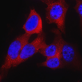
- PKM2 in HeLa Human Cell Line. PKM2 was detected in immersion fixed HeLa human cervical epithelial carcinoma cell line using Mouse Anti-Human/Mouse/Rat PKM2 Monoclonal Antibody (Catalog # MAB72441) at 25 µg/mL for 3 hours at room temperature. Cells were stained using the NorthernLights™ 557-conjugated Anti-Mouse IgG Secondary Antibody (red; Catalog # NL007) and counterstained with DAPI (blue). Specific staining was localized to cytoplasm. View our protocol for Fluorescent ICC Staining of Cells on Coverslips.