Explore
 Validate
 Learn
LearnPA5-23601
antibody from Invitrogen Antibodies
Targeting: KEAP1
INrf2, KIAA0132, KLHL19, MGC10630, MGC1114, MGC20887, MGC4407, MGC9454
 Western blot
Antibody data
- Antibody Data
- Antigen structure
- References [0]
- Comments [0]
- Validations
- Western blot [3]
- Immunocytochemistry [1]
- Immunohistochemistry [1]
Submit
Validation data
Reference
Comment
Report error
- Product number
- PA5-23601 - Provider product page

- Provider
- Invitrogen Antibodies
- Product name
- KEAP1 Polyclonal Antibody
- Antibody type
- Polyclonal
- Antigen
- Synthetic peptide
- Description
- This antibody is predicted to react with porcine based on sequence homology.
- Reactivity
- Human
- Host
- Rabbit
- Isotype
- IgG
- Vial size
- 400 µL
- Concentration
- 0.4 mg/mL
- Storage
- -20° C, Avoid Freeze/Thaw Cycles
No comments: Submit comment
Supportive validation
- Submitted by
- Invitrogen Antibodies (provider)
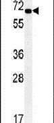
- Main image

- Experimental details
- Western blot analysis using a KEAP1 polyclonal antibody (Product # PA5-23601) in HepG2 cell lysates (35 µg per lane).
- Submitted by
- Invitrogen Antibodies (provider)
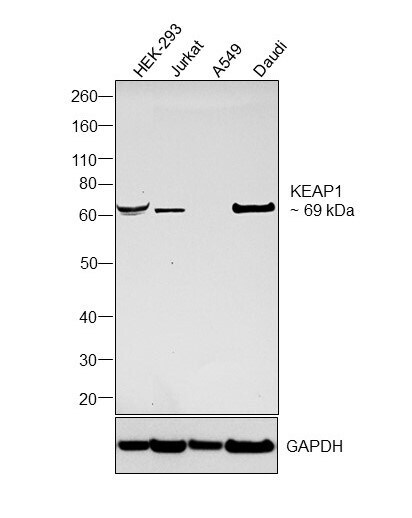
- Main image

- Experimental details
- Western blot was performed using Anti-KEAP1 Polyclonal Antibody (Product # PA5-23601) and a 69 kDa band corresponding to Kelch-like ECH-associated protein 1 was observed across tested cell lines. Whole cell extracts (40 µg lysate) of HEK-293 (Lane 1), Jurkat (Lane 2), A549 (Lane 3), Daudi (Lane 4) were electrophoresed using NuPAGE™ 4-12% Bis-Tris Protein Gel (Product # NP0321BOX). Resolved proteins were then transferred onto a nitrocellulose membrane (Product # IB23001) by iBlot® 2 Dry Blotting System (Product # IB21001). The blot was probed with the primary antibody (1:1000 dilution) and detected by chemiluminescence with Goat anti-Rabbit IgG (H+L) Superclonal™ Recombinant Secondary Antibody, HRP (Product # A27036,1:20000 dilution) using the iBright FL 1000 (Product # A32752). Chemiluminescent detection was performed using SuperSignal™ West Pico PLUS Chemiluminescent Substrate (Product # 34580).
- Submitted by
- Invitrogen Antibodies (provider)
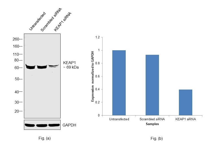
- Main image

- Experimental details
- Knockdown of Kelch-like ECH-associated protein 1 was achieved by transfecting HEK-293 with Kelch-like ECH-associated protein 1 specific siRNAs (Silencer® select Product # s18981, s18982). Western blot analysis (Fig. a) was performed using Whole cell extracts from the Kelch-like ECH-associated protein 1 knockdown cells (lane 3), non-targeting scrambled siRNA transfected cells (lane 2) and untransfected cells (lane 1). The blot was probed with KEAP1 Polyclonal Antibody (Product # PA5-23601, 1:1000 dilution) and Goat anti-Rabbit IgG (H+L) Superclonal™ Recombinant Secondary Antibody, HRP (Product # A27036, 1:20000 dilution). Densitometric analysis of this western blot is shown in histogram (Fig. b). Decrease in signal upon siRNA mediated knock down confirms that antibody is specific to Kelch-like ECH-associated protein 1.
Supportive validation
- Submitted by
- Invitrogen Antibodies (provider)
- Main image
- Experimental details
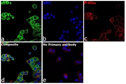
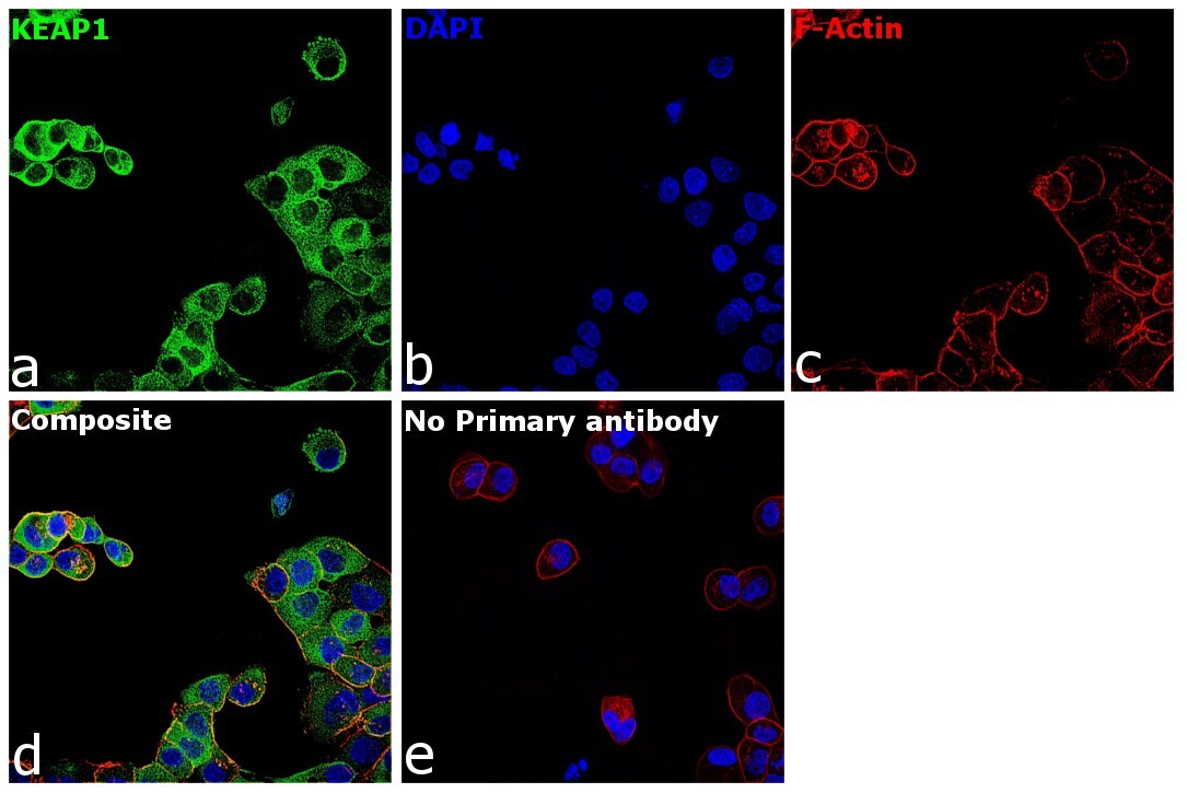
- Immunofluorescence analysis of Kelch-like ECH-associated protein 1 was performed using 70% confluent log phase A-431 cells. The cells were fixed with 4% paraformaldehyde for 5 minutes, permeabilized with 0.01% Triton™ X-100 for 10 minutes, and blocked with 2% BSA for 1 hour at room temperature. The cells were labeled with KEAP1 Polyclonal Antibody (Product # PA5-23601) at 1:100 dilution in 0.1% BSA, incubated at 4 degree celsius overnight and then labeled with Goat anti-Rabbit IgG (H+L) Superclonal™ Recombinant Secondary Antibody, Alexa Fluor® 488 conjugate (Product # A27034), (1:2000 dilution), for 45 minutes at room temperature (Panel a: Green). Nuclei (Panel b:Blue) were stained with Hoechst 33342 (Product # H1399). F-actin (Panel c: Red) was stained with Rhodamine Phalloidin (Product # R415, 1:300). Panel d represents the merged image showing cytoplasm localization. Panel e represents control cells with no primary antibody to assess background. The images were captured at 40X magnification in CellInsight CX7 LZR High-Content Screening (HCS) Platform (Product # CX7A1110LZR) and externally deconvoluted (D.Sage et al. / Methods 115 (2017) 28–41).
Supportive validation
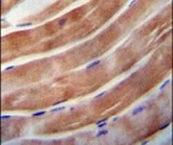
- Submitted by
- Invitrogen Antibodies (provider)
- Main image

- Experimental details
- Immunohistochemistry analysis in formalin-fixed, paraffin-embedded human skeletal muscle using a KEAP1 polyclonal antibody (Product # PA5-23601), followed by HRP-conjugated secondary antibody and DAB staining.