Explore
 Validate
 Learn
LearnPA5-80792
antibody from Invitrogen Antibodies
Targeting: MAPK12
ERK6, p38gamma, PRKM12, SAPK-3, SAPK3
 Western blot
Antibody data
- Antibody Data
- Antigen structure
- References [0]
- Comments [0]
- Validations
- Western blot [3]
- Other assay [1]
Submit
Validation data
Reference
Comment
Report error
- Product number
- PA5-80792 - Provider product page

- Provider
- Invitrogen Antibodies
- Product name
- p38 MAPK gamma Polyclonal Antibody
- Antibody type
- Polyclonal
- Antigen
- Recombinant full-length protein
- Description
- This product is preservative free. It is recommended to add sodium azide to avoid contamination (final concentration 0.05%-0.1%).
- Concentration
- 1 mg/mL
No comments: Submit comment
Supportive validation
- Submitted by
- Invitrogen Antibodies (provider)
- Main image
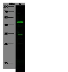
- Experimental details
- Western blot analysis of p38 MAPK gamma in Lane A: Jurkat Whole Cell Lysate (30 µg). Samples were probed using a p38 MAPK gamma Polyclonal Antibody (Product # PA5-80792) at a 1:500 dilution, followed by a Goat Anti-Rabbit IgG (H+L), Dylight 800 Secondary Antibody at a 1:10000 dilution. Western blot was performed under reducing conditions. Predicted band size:42 kDa. Observed band size:45 kDa.

- Submitted by
- Invitrogen Antibodies (provider)
- Main image
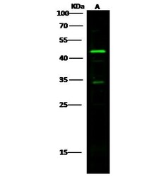
- Experimental details
- Western Blot using p38 MAPK gamma Polyclonal Antibody (Product # PA5-80792) at 1:500 dilution. Lane A: Jurkat Whole Cell Lysate. Lysates/proteins at 30 μg per lane. Secondary Goat Anti-Rabbit IgG H&L (DyLight™ 800) at 1:10,000 dilution. Developed using the Odyssey technique. Performed under reducing conditions. Predicted band size: 42 kDa. Observed band size: 45 kDa. (We are unsure of the identity of these extra bands).

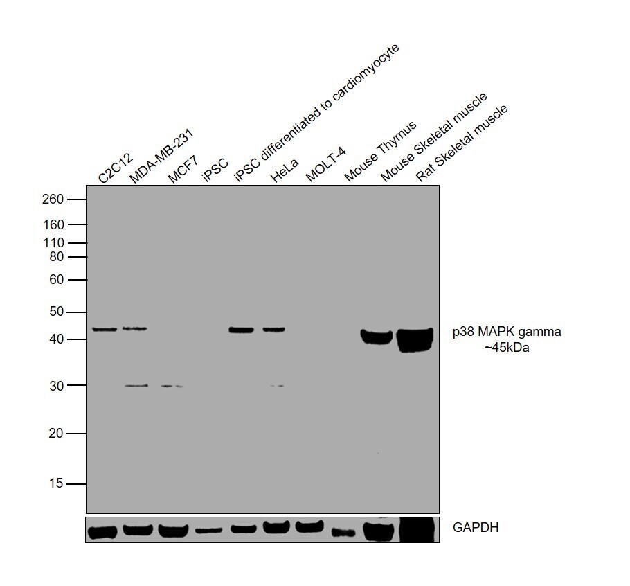
- Submitted by
- Invitrogen Antibodies (provider)
- Main image

- Experimental details
- Western blot was performed using Anti-p38 MAPK gamma Polyclonal Antibody (Product # PA5-80792) and a 45kDa band corresponding to Mitogen-activated protein kinase 12 was observed across all the tested cell lines and tissues except MCF7, MOLT-4 and Mouse Thymus, and was upregulated upon differentiation of iPSC to cardiomyocytes. Whole cell extracts (30 µg lysate) of C2C12 (Lane 1), MDA-MB-231 (Lane 2), MCF7 (Lane 3), iPSC (Lane 4), iPSC (differentiated to cardiomyocytes) (Lane 5), HeLa (Lane 6), MOLT-4 (Lane 7), Mouse Thymus (Lane 8), Mouse Skeletal Muscle (Lane 9), Rat Skeletal Muscle (Lane 10) were electrophoresed using NuPAGE™ 10% Bis-Tris Protein Gel (Product # NP0302BOX). Resolved proteins were then transferred onto a Nitrocellulose membrane (Product # IB23001) by iBlot® 2 Dry Blotting System (Product # IB21001). The blot was probed with the primary antibody (1:1000 dilution) and detected by chemiluminescence with Goat anti-Rabbit IgG (H+L) Superclonal™ Recombinant Secondary Antibody, HRP (Product # A27036, 1:4000 dilution) using the iBright FL 1000 (Product # A32752). Chemiluminescent detection was performed using Novex® ECL Reagent Kit (Product # WP20005).
Supportive validation
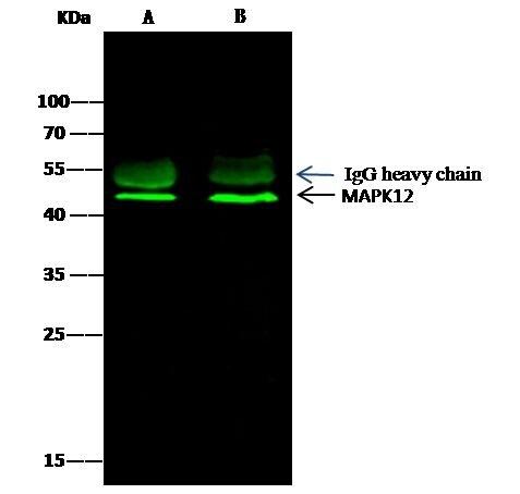
- Submitted by
- Invitrogen Antibodies (provider)
- Main image
- Experimental details
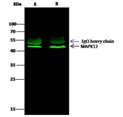
- p38 MAPK gamma Immunoprecipitation using: Lane A: 0.5 mg Hela Whole Cell Lysate, Lane B: 0.5 mg 293T Whole Cell Lysate 0.5 µL with p38 MAPK gamma Polyclonal Antibody (Product # PA5-80792) and 15 µL of 50 % Protein G agarose. Primary antibody: p38 MAPK gamma Polyclonal Antibody, at 1:500 dilution. Secondary antibody: Dylight 800-labeled antibody to rabbit IgG (H+L), at 1:5,000 dilution. Developed using the Odyssey technique. Performed under reducing conditions. Predicted band size: 42 kDa. Observed band size: 42 kDa.