Explore
 Validate
 Learn
Learn Flow cytometry
Antibody data
- Antibody Data
- Antigen structure
- References [8]
- Comments [0]
- Validations
- Flow cytometry [1]
- Other assay [14]
Submit
Validation data
Reference
Comment
Report error
- Product number
- 13-8888-82 - Provider product page

- Provider
- Invitrogen Antibodies
- Product name
- CD338 (ABCG2) Monoclonal Antibody (5D3), Biotin, eBioscience™
- Antibody type
- Monoclonal
- Antigen
- Other
- Description
- Description: The 5D3 monoclonal antibody reacts with the extracellular portion of the human ABCG2 protein, also known as Bcrp1 and MXR. The ABCG2 gene, a member of the multi-drug resistance (MDR) family, is highly expressed on primitive 'side-population' (SP) stem cells, which are defined by the efflux of fluorescent dyes such as Rhodamine 123 and Hoechest 33342. In the bone marrow, about 0.05% of cells display the low fluorescence and are highly enriched for repopulating cells. The SP cells, which express low or undetectable levels of CD34, have been identified in multiple species. In addition, expression of ABCG2 appears to be highly conserved.
- Conjugate
- Biotin
- Antibody clone number
- 5D3
- Concentration
- 0.5 mg/mL
Submitted references CBP-mediated Wnt3a/β-catenin signaling promotes cervical oncogenesis initiated by Piwil2.
Piwil2 is reactivated by HPV oncoproteins and initiates cell reprogramming via epigenetic regulation during cervical cancer tumorigenesis.
ABCG2pos lung mesenchymal stem cells are a novel pericyte subpopulation that contributes to fibrotic remodeling.
Human prostate side population cells demonstrate stem cell properties in recombination with urogenital sinus mesenchyme.
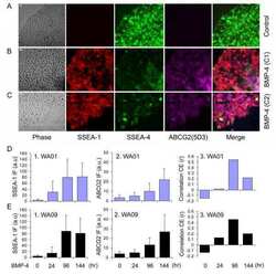
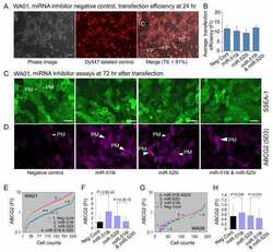
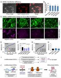
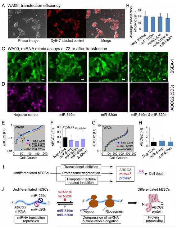
Regulation and expression of the ATP-binding cassette transporter ABCG2 in human embryonic stem cells.
The PI3K/Akt inhibitor LY294002 reverses BCRP-mediated drug resistance without affecting BCRP translocation.
Histone deacetylase inhibitors influence chemotherapy transport by modulating expression and trafficking of a common polymorphic variant of the ABCG2 efflux transporter.
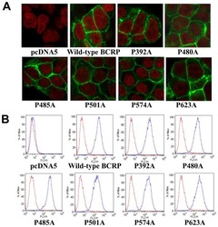
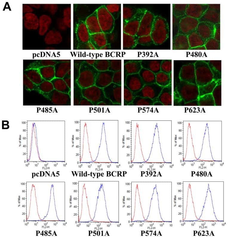
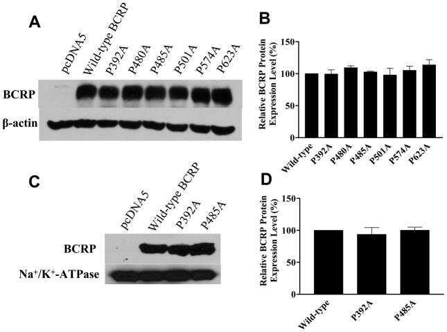
Identification of proline residues in or near the transmembrane helices of the human breast cancer resistance protein (BCRP/ABCG2) that are important for transport activity and substrate specificity.
Feng D, Yan K, Liang H, Liang J, Wang W, Yu H, Zhou Y, Zhao W, Dong Z, Ling B
Neoplasia (New York, N.Y.) 2021 Jan;23(1):1-11
Neoplasia (New York, N.Y.) 2021 Jan;23(1):1-11
Piwil2 is reactivated by HPV oncoproteins and initiates cell reprogramming via epigenetic regulation during cervical cancer tumorigenesis.
Feng D, Yan K, Zhou Y, Liang H, Liang J, Zhao W, Dong Z, Ling B
Oncotarget 2016 Oct 4;7(40):64575-64588
Oncotarget 2016 Oct 4;7(40):64575-64588
ABCG2pos lung mesenchymal stem cells are a novel pericyte subpopulation that contributes to fibrotic remodeling.
Marriott S, Baskir RS, Gaskill C, Menon S, Carrier EJ, Williams J, Talati M, Helm K, Alford CE, Kropski JA, Loyd J, Wheeler L, Johnson J, Austin E, Nozik-Grayck E, Meyrick B, West JD, Klemm DJ, Majka SM
American journal of physiology. Cell physiology 2014 Oct 15;307(8):C684-98
American journal of physiology. Cell physiology 2014 Oct 15;307(8):C684-98
Human prostate side population cells demonstrate stem cell properties in recombination with urogenital sinus mesenchyme.
Foster BA, Gangavarapu KJ, Mathew G, Azabdaftari G, Morrison CD, Miller A, Huss WJ
PloS one 2013;8(1):e55062
PloS one 2013;8(1):e55062
Regulation and expression of the ATP-binding cassette transporter ABCG2 in human embryonic stem cells.
Padmanabhan R, Chen KG, Gillet JP, Handley M, Mallon BS, Hamilton RS, Park K, Varma S, Mehaffey MG, Robey PG, McKay RD, Gottesman MM
Stem cells (Dayton, Ohio) 2012 Oct;30(10):2175-87
Stem cells (Dayton, Ohio) 2012 Oct;30(10):2175-87
The PI3K/Akt inhibitor LY294002 reverses BCRP-mediated drug resistance without affecting BCRP translocation.
Imai Y, Yoshimori M, Fukuda K, Yamagishi H, Ueda Y
Oncology reports 2012 Jun;27(6):1703-9
Oncology reports 2012 Jun;27(6):1703-9
Histone deacetylase inhibitors influence chemotherapy transport by modulating expression and trafficking of a common polymorphic variant of the ABCG2 efflux transporter.
Basseville A, Tamaki A, Ierano C, Trostel S, Ward Y, Robey RW, Hegde RS, Bates SE
Cancer research 2012 Jul 15;72(14):3642-51
Cancer research 2012 Jul 15;72(14):3642-51
Identification of proline residues in or near the transmembrane helices of the human breast cancer resistance protein (BCRP/ABCG2) that are important for transport activity and substrate specificity.
Ni Z, Bikadi Z, Shuster DL, Zhao C, Rosenberg MF, Mao Q
Biochemistry 2011 Sep 20;50(37):8057-66
Biochemistry 2011 Sep 20;50(37):8057-66
No comments: Submit comment
Supportive validation
- Submitted by
- Invitrogen Antibodies (provider)
- Main image

- Experimental details
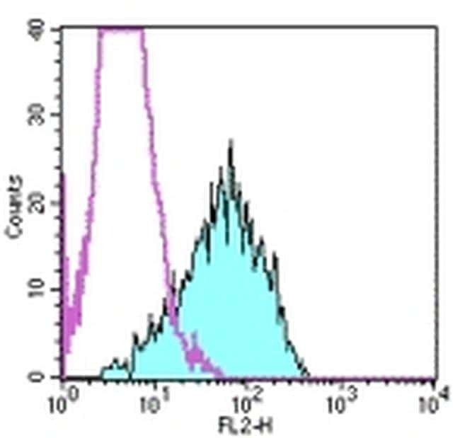
- Staining of MCF/huBCRP-1 transfected cells with 0.5 µg of Anti-Mouse IgG2b Biotin (Product # 13-4732-85) (open histogram) or 0.5 µg of Anti-Human CD338 (ABCG2) Biotin followed by Streptavidin PE (Product # 12-4317-87) (filled histogram). Total viable cells were used for analysis.
- Conjugate
- Biotin
Supportive validation
- Submitted by
- Invitrogen Antibodies (provider)
- Main image
- Experimental details
- NULL
- Conjugate
- Biotin
- Submitted by
- Invitrogen Antibodies (provider)
- Main image
- Experimental details
- NULL
- Conjugate
- Biotin
- Submitted by
- Invitrogen Antibodies (provider)
- Main image

- Experimental details
- NULL
- Conjugate
- Biotin
- Submitted by
- Invitrogen Antibodies (provider)
- Main image

- Experimental details
- NULL
- Conjugate
- Biotin
- Submitted by
- Invitrogen Antibodies (provider)
- Main image
- Experimental details
- NULL
- Conjugate
- Biotin

- Submitted by
- Invitrogen Antibodies (provider)
- Main image
- Experimental details
- NULL
- Conjugate
- Biotin

- Submitted by
- Invitrogen Antibodies (provider)
- Main image
- Experimental details
- NULL
- Conjugate
- Biotin
- Submitted by
- Invitrogen Antibodies (provider)
- Main image
- Experimental details
- NULL
- Conjugate
- Biotin

- Submitted by
- Invitrogen Antibodies (provider)
- Main image

- Experimental details
- NULL
- Conjugate
- Biotin
- Submitted by
- Invitrogen Antibodies (provider)
- Main image

- Experimental details
- NULL
- Conjugate
- Biotin
- Submitted by
- Invitrogen Antibodies (provider)
- Main image
- Experimental details
- NULL
- Conjugate
- Biotin

- Submitted by
- Invitrogen Antibodies (provider)
- Main image
- Experimental details
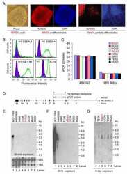
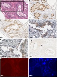
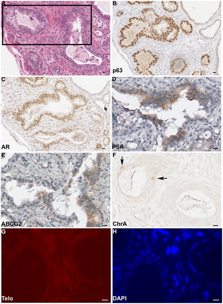
- Figure 4 Expression of Differentiation Markers in Recombinants. Nonconsecutive serial sections of a recombinant derived from 250 side population cells from specimen NT6+ rUGM were stained. A) H&E staining, box represents area examined at higher power in D-H; B) p63 IHC; C) AR IHC; D) PSA IHC; E) ABCG2 IHC; F) Chromogranin A (ChrA) IHC arrows indicate positive cells; G) telomere (Telo) detection by FISH; and H) DAPI staining. B-H) Scale bar, 10 um.
- Conjugate
- Biotin
- Submitted by
- Invitrogen Antibodies (provider)
- Main image
- Experimental details
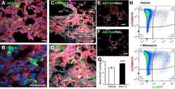
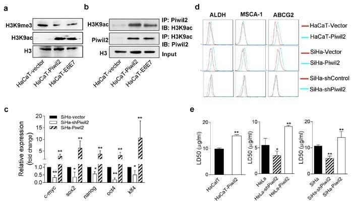
- Figure 7 Piwil2 initiates cell reprogramming by regulating the balance between the acetylation and trimethylation of H3K9 a. Western blot showing significantly induced H3K9 acetylation but reduced H3K9 trimethylation in HaCaT cells transfected with Piwil2 or E6 and E7 compared with those only transfected with vector. b. Co-immunoprecipitation showing that Piwil2, either overexpressed or induced by E6 and E7, interacted with acetylated H3K9. c. EMT markers upregulated in SiHa cells exhibiting Piwil2 overexpression but downregulated in those cells in which Piwil2 was knocked down, as verified by qRT-PCR. d. The proportion of ALDH-, MSCA-1-, and ABCG2-positive cells, determined by FACS in cells with Piwil2 overexpression or knockdown. e. The LD50 dose of cisplatin, detected by CCK8 assay in cells with Piwil2 overexpression or knockdown. The data are presented as the mean+-SD. * P < 0.05 and ** P < 0.01 by Student's t -test.
- Conjugate
- Biotin
- Submitted by
- Invitrogen Antibodies (provider)
- Main image
- Experimental details
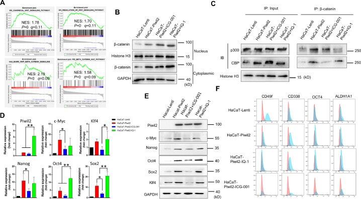
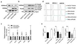
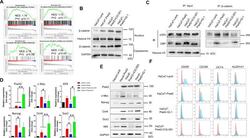
- Figure 4 CBP/beta-catenin promotes the maintenance of stem cell reprogramming by Piwil2. (A) GSEA plot showing significant enrichment of the Wnt/beta-catenin signaling activation modules in HaCaT-Piwil2 cells. (B) Immunoblotting of beta-catenin translocated into the nucleus in HaCaT-Piwil2 cells after treatment with 20 uM ICG-001 or IQ-1 for 24 h. (C) Nuclear lysates from HaCaT-Piwil2 cells treated with 20 uM ICG-001, 20 uM IQ-1, or DMSO were coimmunoprecipitated with antisera to beta-catenin and immunoblotted for CBP and p300. (D and E) The expression of Piwil2 and the ""reprogramming"" factors c-Myc, Nanog, Oct4, Sox2 , and Klf4 was determined by real-time PCR and immunoblotting in HaCaT-Piwil2 cells treated with 20 uM ICG-001, 20 uM IQ-1, or DMSO for 24 h. (F) The proportion of CD49f-, CD338-, OCT4-, and ALDHA1-positive cells, determined by flow cytometry in HaCaT-Piwil2 cells treated with 20 uM ICG-001, 20 uM IQ-1, or DMSO for 24 h. The data are presented as the mean +- SD. * P < 0.05 and ** P < 0.01 by Student's t test. Figure 4
- Conjugate
- Biotin