Explore
 Validate
 Learn
Learn Other assay
Antibody data
- Antibody Data
- Antigen structure
- References [3]
- Comments [0]
- Validations
- Other assay [2]
Submit
Validation data
Reference
Comment
Report error
- Product number
- MA1-20167 - Provider product page

- Provider
- Invitrogen Antibodies
- Product name
- NGFR Monoclonal Antibody (ME20.4)
- Antibody type
- Monoclonal
- Antigen
- Other
- Description
- Recommended positive controls: human tongue.
- Antibody clone number
- ME20.4
- Concentration
- 2.1 mg/mL
Submitted references Development of a functional schwann cell phenotype from autologous porcine bone marrow mononuclear cells for nerve repair.
Functional sphere profiling reveals the complexity of neuroblastoma tumor-initiating cell model.
Functional sphere profiling reveals the complexity of neuroblastoma tumor-initiating cell model.
Rutten MJ, Janes MA, Chang IR, Gregory CR, Gregory KW
Stem cells international 2012;2012:738484
Stem cells international 2012;2012:738484
Functional sphere profiling reveals the complexity of neuroblastoma tumor-initiating cell model.
Coulon A, Flahaut M, Mühlethaler-Mottet A, Meier R, Liberman J, Balmas-Bourloud K, Nardou K, Yan P, Tercier S, Joseph JM, Sommer L, Gross N
Neoplasia (New York, N.Y.) 2011 Oct;13(10):991-1004
Neoplasia (New York, N.Y.) 2011 Oct;13(10):991-1004
No comments: Submit comment
Supportive validation
- Submitted by
- Invitrogen Antibodies (provider)
- Main image
- Experimental details
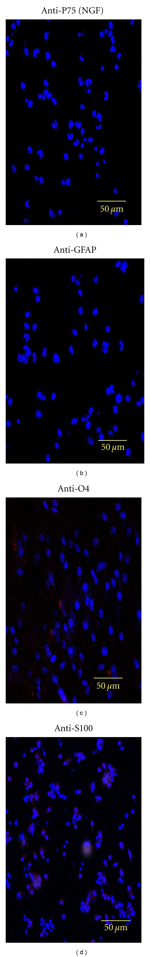
- Figure 3 Series of confocal photographs showing that freshly isolated porcine BM-MNCs do not express Schwann cell markers in neurobasal media. The cells were found to be negative staining for the Schwann cell markers p75(NGF), GFAP, O4, and S100 (a, b, c, d). Nuclei (blue) are stained with DAPI.
- Submitted by
- Invitrogen Antibodies (provider)
- Main image
- Experimental details
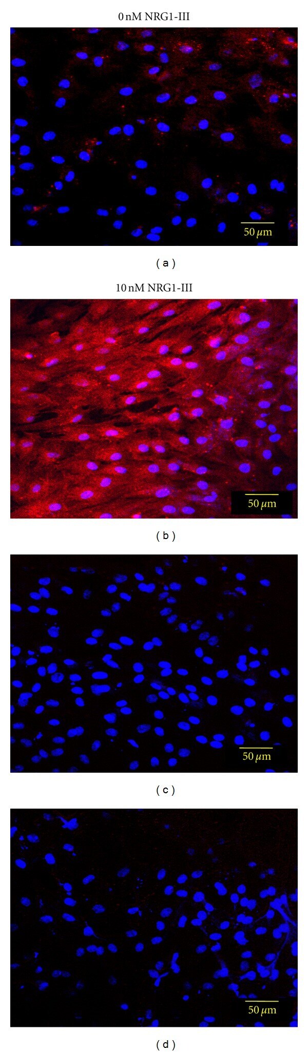
- Figure 5 Confocal photographs showing that BM-MNCs cultured in neurobasal media supplemented with NRG1-III express the p75(NGF) receptor. The addition of 10 nM NRG1-III for 48-hrs to 6-day-old cultures increased the expression of p75(NGF) (b) over untreated control cells (a); (c, d) are control cultures stained with only the secondary antibody. The nuclei (blue) were stained with DAPI.