Explore
 Validate
 Learn
Learn Western blot
Antibody data
- Antibody Data
- Antigen structure
- References [0]
- Comments [0]
- Validations
- Western blot [1]
- Immunocytochemistry [2]
- Immunohistochemistry [13]
- Flow cytometry [3]
Submit
Validation data
Reference
Comment
Report error
- Product number
- GTX84135 - Provider product page

- Provider
- GeneTex
- Proper citation
- GeneTex Cat#GTX84135, RRID:AB_10730335
- Product name
- MCL1 antibody [2E11]
- Antibody type
- Monoclonal
- Reactivity
- Human, Simian
- Host
- Mouse
No comments: Submit comment
Supportive validation
- Submitted by
- GeneTex (provider)
- Main image
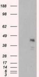
- Experimental details
- HEK293T cells were transfected with the pCMV6-ENTRY control (Left lane) or pCMV6-ENTRY MCL1 (Right lane) cDNA for 48 hrs and lysed. Equivalent amounts of cell lysates (5 ug per lane) were separated by SDS-PAGE and immunoblotted with anti-MCL1.
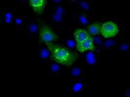
Supportive validation
- Submitted by
- GeneTex (provider)
- Main image

- Experimental details
- Anti-MCL1 mouse monoclonal antibody (GTX84135) immunofluorescent staining of COS7 cells transiently transfected with MCL1
- Submitted by
- GeneTex (provider)
- Main image
- Experimental details
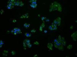
- Immunofluorescent staining of HepG2 cells using anti-MCL1 mouse monoclonal antibody (GTX84135).

Supportive validation
- Submitted by
- GeneTex (provider)
- Main image
- Experimental details
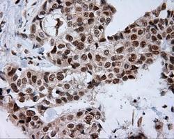
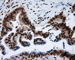
- Immunohistochemical staining of paraffin-embedded Adenocarcinoma of breast tissue using anti-MCL1 mouse monoclonal antibody. (GTX84135, Dilution 1:50)

- Submitted by
- GeneTex (provider)
- Main image

- Experimental details
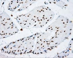
- Immunohistochemical staining of paraffin-embedded lung tissue using anti-MCL1 mouse monoclonal antibody. (GTX84135, Dilution 1:50)

- Submitted by
- GeneTex (provider)
- Main image
- Experimental details
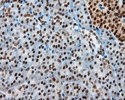
- Immunohistochemical staining of paraffin-embedded pancreas tissue using anti-MCL1 mouse monoclonal antibody. (GTX84135, Dilution 1:50)

- Submitted by
- GeneTex (provider)
- Main image
- Experimental details
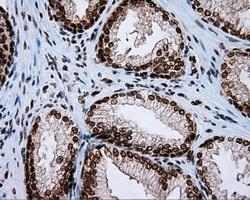
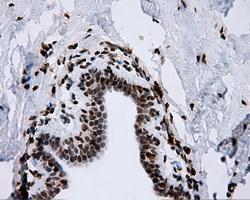
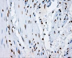
- Immunohistochemical staining of paraffin-embedded prostate tissue using anti-MCL1 mouse monoclonal antibody. (GTX84135, Dilution 1:50)

- Submitted by
- GeneTex (provider)
- Main image
- Experimental details
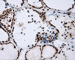
- Immunohistochemical staining of paraffin-embedded thyroid tissue using anti-MCL1 mouse monoclonal antibody. (GTX84135, Dilution 1:50)

- Submitted by
- GeneTex (provider)
- Main image

- Experimental details
- Immunohistochemical staining of paraffin-embedded breast tissue using anti-MCL1 mouse monoclonal antibody. (GTX84135, Dilution 1:50)
- Submitted by
- GeneTex (provider)
- Main image

- Experimental details
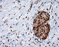
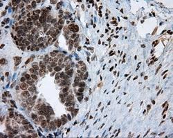
- Immunohistochemical staining of paraffin-embedded Carcinoma of pancreas tissue using anti-MCL1 mouse monoclonal antibody. (GTX84135, Dilution 1:50)
- Submitted by
- GeneTex (provider)
- Main image
- Experimental details
- Immunohistochemical staining of paraffin-embedded Carcinoma of thyroid tissue using anti-MCL1 mouse monoclonal antibody. (GTX84135, Dilution 1:50)

- Submitted by
- GeneTex (provider)
- Main image
- Experimental details
- Immunohistochemical staining of paraffin-embedded Adenocarcinoma of colon tissue using anti-MCL1 mouse monoclonal antibody. (GTX84135, Dilution 1:50)

- Submitted by
- GeneTex (provider)
- Main image

- Experimental details
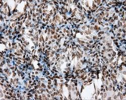
- Immunohistochemical staining of paraffin-embedded Adenocarcinoma of ovary tissue using anti-MCL1 mouse monoclonal antibody. (GTX84135, Dilution 1:50)
- Submitted by
- GeneTex (provider)
- Main image
- Experimental details
- Immunohistochemical staining of paraffin-embedded bladder tissue using anti-MCL1 mouse monoclonal antibody. (GTX84135, Dilution 1:50)

- Submitted by
- GeneTex (provider)
- Main image
- Experimental details
- Immunohistochemical staining of paraffin-embedded colon tissue using anti-MCL1 mouse monoclonal antibody. (GTX84135, Dilution 1:50)

- Submitted by
- GeneTex (provider)
- Main image

- Experimental details
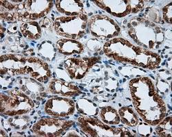
- Immunohistochemical staining of paraffin-embedded Kidney tissue using anti-MCL1 mouse monoclonal antibody. (GTX84135, Dilution 1:50)
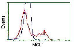
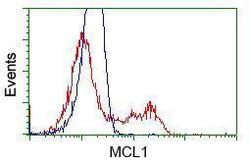
Supportive validation
- Submitted by
- GeneTex (provider)
- Main image
- Experimental details
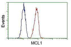
- Flow cytometric analysis of Hela cells, using anti-MCL1 antibody(GTX84135),(Red) compared to a nonspecific negative control antibody(TA50011)(Blue).
- Submitted by
- GeneTex (provider)
- Main image
- Experimental details
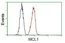
- Flow cytometric analysis of Jurkat cells, using anti-MCL1 antibody(GTX84135),(Red) compared to a nonspecific negative control antibody(TA50011)(Blue).

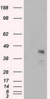
- Submitted by
- GeneTex (provider)
- Main image
- Experimental details
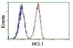
- HEK293T cells transfected with either pCMV6-ENTRY MCL1