Explore
 Validate
 Learn
Learn Flow cytometry
Antibody data
- Antibody Data
- Antigen structure
- References [0]
- Comments [0]
- Validations
- Flow cytometry [1]
Submit
Validation data
Reference
Comment
Report error
- Product number
- 12-9038-42 - Provider product page

- Provider
- Invitrogen Antibodies
- Product name
- Phospho-MCL-1 (Ser159) Monoclonal Antibody (RBCERNR), PE, eBioscience™
- Antibody type
- Monoclonal
- Antigen
- Other
- Description
- Description: This RBCERNR monoclonal antibody recognizes human and mouse myeloid cell leukemia sequence 1 (Mcl-1) when phosphorylated on serine 159 (S159). Mcl-1 is an anti-apoptotic protein that is a member of the Bcl-2 family of proteins important for regulation of cell survival/apoptosis. Mcl-1 is primarily localized to the outer membrane of mitochondria where it prevents cytochrome c release via dimerization with other Bcl-2 family members such as Bim. PI3K activation of AKT results in the phosphorylation of GSK3 beta at serine 9 (S9) resulting in destabilization and degradation of GSK3 beta. Loss of GSK3 beta prevents phosphorylation of Mcl-1 on S159 and its subsequent ubiquitnation and degradation. Mice conditionally lacking Mcl-1 in lymphocytes showed that Mcl-1 is essential during early lymphoid development and for the maintenance of mature lymphocytes.
- Conjugate
- Yellow dye
- Antibody clone number
- RBCERNR
- Concentration
- 5 µL/Test
No comments: Submit comment
Supportive validation
- Submitted by
- Invitrogen Antibodies (provider)
- Main image

- Experimental details
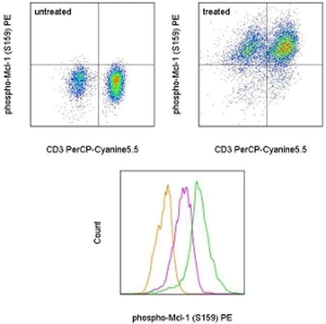
- TOP: Intracellular staining of normal human peripheral blood cells that were untreated (left) or treated with Calyculin A for 4 hours (right) with Anti-Human CD3 PerCP-Cyanine5-5 (Product # 45-0036-42) and phospho-Mcl-1 (S159) PE. Plots show cells in the lymphocyte gate. BOTTOM: Normal human peripheral blood cells were unstimulated (orange histogram), were stimulated with Anti-Human CD3 and CD28 Functional Grade Purifieds (Product # 16-0037-81 and Product # 16-0289-81) in the presence of the proteasome inhibitor MG-132 (purple histogram), or were treated with Calyculin A (green histogram). The cells were then intracellularly stained with Anti-Human CD3 PerCP-Cyanine5-5 (Product # 45-0036-42) and Anti-Human/Mouse phospho-Mcl-1 (S159) PE using the Intracellular Fixation & Permeabilization Buffer Set (Product # 88-8824-00) and protocol. CD3+ cells in the lymphocyte gate were used for analysis.
- Conjugate
- Yellow dye