Explore
 Validate
 Learn
Learn Western blot
Antibody data
- Antibody Data
- Antigen structure
- References [0]
- Comments [0]
- Validations
- Western blot [6]
- Immunocytochemistry [1]
- Flow cytometry [1]
Submit
Validation data
Reference
Comment
Report error
- Product number
- MA5-17084 - Provider product page

- Provider
- Invitrogen Antibodies
- Product name
- GluR2 Monoclonal Antibody (7G6)
- Antibody type
- Monoclonal
- Antigen
- Purifed from natural sources
- Description
- MA5-17084 targets GRIA2 in FACS, pep-ELISA, and WB applications and shows reactivity with Human samples.
- Antibody clone number
- 7G6
- Concentration
- 1 mg/mL
No comments: Submit comment
Supportive validation
- Submitted by
- Invitrogen Antibodies (provider)
- Main image
- Experimental details
- Western blot analysis of GRIA2 using a GRIA2 monoclonal antibody (Product # MA5-17084) against a human GRIA2 recombinant protein.

- Submitted by
- Invitrogen Antibodies (provider)
- Main image

- Experimental details
- Western blot analysis of GRIA2 using GRIA2 monoclonal antibody (Product # MA5-17084) in HEK293 (1) and GRIA2 (AA: 652-807) human IgG Fc transfected HEK293 (2) cell lysate.
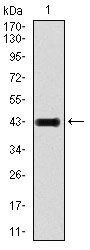
- Submitted by
- Invitrogen Antibodies (provider)
- Main image

- Experimental details
- Western blot analysis of GRIA2 using a GRIA2 monoclonal antibody (Product # MA5-17084) against a human GRIA2 recombinant protein.

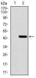
- Submitted by
- Invitrogen Antibodies (provider)
- Main image

- Experimental details
- Western blot analysis of GRIA2 using GRIA2 monoclonal antibody (Product # MA5-17084) in HEK293 (1) and GRIA2 (AA: 652-807) human IgG Fc transfected HEK293 (2) cell lysate.

- Submitted by
- Invitrogen Antibodies (provider)
- Main image
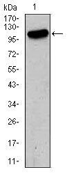
- Experimental details
- Western blot analysis of GRIA2 using GRIA2 monoclonal antibody (Product # MA5-17084) in HeLa (1) cell lysate.

- Submitted by
- Invitrogen Antibodies (provider)
- Main image
- Experimental details
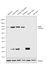
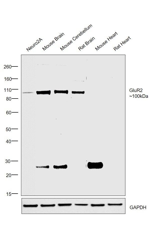
- Western blot was performed using Anti-GluR2 Monoclonal Antibody (7G6) (Product # MA5-17084) and a 100kDa band corresponding to GluR2 was observed across all cell lines and tissues tested except Mouse Heart and Rat Heart which is reported to be negative. Membrane enriched cell extracts (30 µg lysate) of Neuro2A (Lane 1), Mouse Brain (Lane 2), Mouse Cerebellum (Lane 3), Rat Brain (Lane 4), Mouse Heart (Lane 5) and Rat Heart (Lane 6) were electrophoresed using Novex® NuPAGE® 4-12 % Bis-Tris gel (Product # NP0322BOX). Resolved proteins were then transferred onto a nitrocellulose membrane (Product # IB23001) by iBlot® 2 Dry Blotting System (Product # IB21001). The blot was probed with the primary antibody (1:1000 dilution) and detected by chemiluminescence with Goat anti-Mouse IgG (H+L), Superclonal™ Recombinant Secondary Antibody, HRP (Product # A28177, 1:4000 dilution) using the iBright FL 1000 (Product # A32752). Chemiluminescent detection was performed using Novex® ECL Chemiluminescent Substrate Reagent Kit (Product # WP20005). A ~25kDa band corresponding to circulating tissue IgG was observed in case of Mouse tissue lysates..
Supportive validation
- Submitted by
- Invitrogen Antibodies (provider)
- Main image
- Experimental details
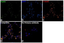
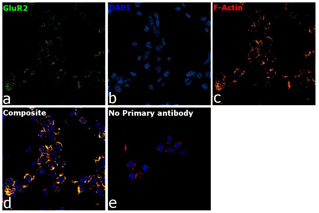
- Immunofluorescence analysis of GluR2 Monoclonal Antibody (7G6) was performed using 70% confluent log phase Neuro-2a cells. The cells were fixed with 4% Paraformaldehyde for 10 minutes, permeabilized with 0.1% Triton™ X-100 for 10 minutes, and blocked with 2% BSA for 10 minutes at room temperature. The cells were labeled with GluR2 Monoclonal Antibody (Product # MA5-17084) at 1:100 dilution in 0.1% BSA, incubated at 4 degree celsius overnight and then labeled Goat anti-Mouse IgG (H+L) Superclonal™ Secondary Antibody, Alexa Fluor® 488 conjugate (Product # A28175, 1:2000 dilution) for 45 minutes at room temperature (Panel a: Green). Nuclei (Panel b: Blue) were stained with SlowFade® Gold Antifade Mountant with DAPI (Product # S36938). F-actin (Panel c: Red) was stained with Rhodamine Phalloidin (Product # R415, 1:300). Panel d represents the merged image showing membrane localization. Panel e represents control cells with no primary antibody to assess background. The images were captured at 60X magnification..
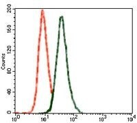
Supportive validation
- Submitted by
- Invitrogen Antibodies (provider)
- Main image
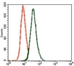
- Experimental details
- Flow cytometric analysis of SK-N-SH cells using GRIA2 monoclonal antibody (Product # MA5-17084) (green) and negative control (red).