 Explore
 Validate
 Learn
Learn Western blot
Antibody data
- Antibody Data
- Antigen structure
- References [0]
- Comments [0]
- Validations
- Western blot [3]
- Immunocytochemistry [1]
- Immunohistochemistry [3]
Submit
Validation data
Reference
Comment
Report error
- Product number
- PA5-29033 - Provider product page

- Provider
- Invitrogen Antibodies
- Product name
- Cytokeratin 7 Polyclonal Antibody
- Antibody type
- Polyclonal
- Antigen
- Recombinant protein fragment
- Description
- Recommended positive controls: A431, HeLa, Molt-4.
- Concentration
- 1.64 mg/mL
No comments: Submit comment
Supportive validation
- Submitted by
- Invitrogen Antibodies (provider)
- Main image

- Experimental details
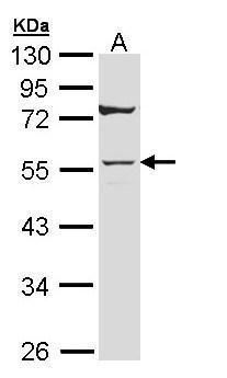
- Western blot analysis of Cytokeratin 7 using 30 µg of A431 lysate. Samples were loaded onto a 10% SDS-PAGE gel and probed with a Cytokeratin 7 polyclonal antibody (Product # PA5-29033) at a dilution of 1:10,000.
- Submitted by
- Invitrogen Antibodies (provider)
- Main image
- Experimental details
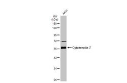
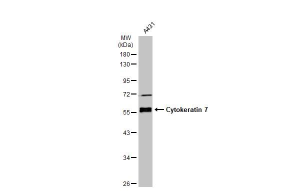
- Western Blot using Cytokeratin 7 Polyclonal Antibody (Product # PA5-29033). Whole cell extract (30 µg) was separated by 10% SDS-PAGE, and the membrane was blotted with Cytokeratin 7 Polyclonal Antibody (Product # PA5-29033) diluted at 1:20,000. The HRP-conjugated anti-rabbit IgG antibody was used to detect the primary antibody.
- Submitted by
- Invitrogen Antibodies (provider)
- Main image

- Experimental details
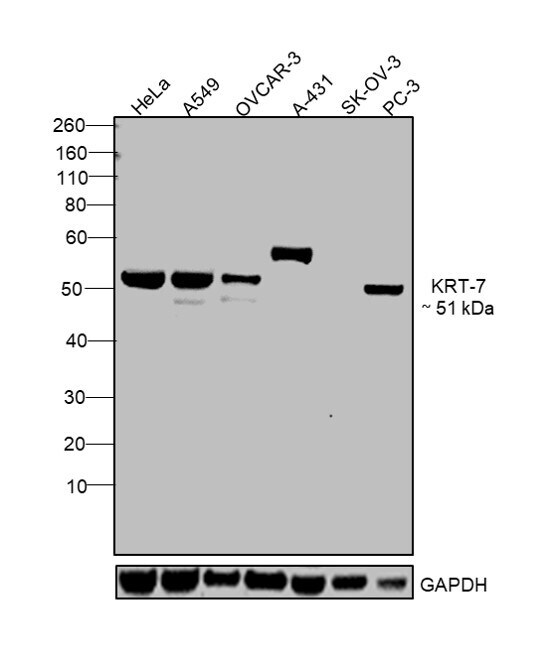
- Western blot was performed using Anti-Cytokeratin 7 Polyclonal Antibody (Product # PA5-29033) and a 51 kDa band corresponding to KRT-7 was observed in positive cell lines like HeLa, A549, OVCAR-3, A-431, PC-3 and not in negative cell lines SK-OV-3 as mentioned in literature. Membrane enriched extracts (30 µg lysate) of HeLa (Lane 1), A549 (Lane 2), OVCAR-3 (Lane 3), A-431 (Lane 4), SK-OV-3 (Lane 5) and PC-3 (Lane 6) were electrophoresed using NuPAGE® 4-12 % Bis-Tris gel (Product # NP0321BOX). Resolved proteins were then transferred onto a nitrocellulose membrane (Product # IB23001) by iBlot® 2 Dry Blotting System (Product # IB21001). The blot was probed with the primary antibody (1:10000 dilution) and detected by chemiluminescence with Goat anti-Rabbit IgG (H+L), Superclonal™ Recombinant Secondary Antibody, HRP conjugate (Product # A27036, 1:4000 dilution) using the iBright FL 1000 (Product # A32752). Chemiluminescent detection was performed using Novex® ECL Chemiluminescent Substrate Reagent Kit (Product # WP20005).
Supportive validation
- Submitted by
- Invitrogen Antibodies (provider)
- Main image

- Experimental details
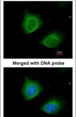
- Immunofluorescent analysis of Cytokeratin 7 in paraformaldehyde-fixed HeLa cells using a Cytokeratin 7 polyclonal antibody (Product # PA5-29033) at a 1:200 dilution.
Supportive validation
- Submitted by
- Invitrogen Antibodies (provider)
- Main image

- Experimental details
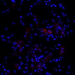
- Immunohistochemistry analysis of Synaptophysin and Cytokeratin 7 was performed in human small cell lung cancer (SCLC) (upper panel) and non-small cell lung cancer (NSCLC) (lower panel) specimens. The section was pre-treated tissue using heat mediated antigen retrieval with sodium citrate buffer (pH6) for 15 mins. The section was then incubated with Synaptophysin Polyclonal Antibody (Product # PA5-27286) or Cytokeratin 7 Polyclonal Antibody (Product # PA5-29033) at 1:500 overnight at 4°C and detected using an HRP conjugated avidin-biotin-peroxidase Complex system. DAB was used as the chromogen and counterstained with haematoxylin. Antigen Retrieval: Citrate buffer, pH 6.0, 15 min.

- Submitted by
- Invitrogen Antibodies (provider)
- Main image
- Experimental details
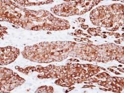
- Immunohistochemical analysis of human placenta (paraformaldehyde-fixed frozen sections), using Cytokeratin 7 (Product # PA5-29033) antibody at 1:200 dilution. Antigen Retrieval: Citrate buffer, pH 6.0, 15 min.

- Submitted by
- Invitrogen Antibodies (provider)
- Main image
- Experimental details
- Immunohistochemical analysis of paraffin-embedded A549 xenograft, using Cytokeratin 7 (Product # PA5-29033) antibody at 1:500 dilution. Antigen Retrieval: EDTA based buffer, pH 8.0, 15 min.
