Explore
 Validate
 Learn
Learn Western blot
 Immunocytochemistry
 Immunohistochemistry
Antibody data
- Antibody Data
- Antigen structure
- References [1]
- Comments [0]
- Validations
- Immunocytochemistry [1]
- Immunohistochemistry [9]
Submit
Validation data
Reference
Comment
Report error
- Product number
- AMAb90599 - Provider product page

- Provider
- Atlas Antibodies
- Proper citation
- Atlas Antibodies Cat#AMAb90599, RRID:AB_2665603
- Product name
- Anti-S100A4
- Antibody type
- Monoclonal
- Reactivity
- Human
- Host
- Mouse
- Conjugate
- Unconjugated
- Antigen sequence
MACPLEKALDVMVSTFHKYSGKEGDKFKLNKSELK
ELLTRELPSFLGKRTDEAAFQKLMSNLDSNRDNEV
DFQEYCVFLSCIAMMCNEFFEGFPDKQPRKK- Epitope
- Binds to an epitope located within the peptide sequence CNEFFEGFPD as determined by overlapping synthetic peptides.
- Isotype
- IgG
- Antibody clone number
- CL0240
- Vial size
- 100 µl
- Storage
- Store at +4°C for short term storage. Long time storage is recommended at -20°C.
Submitted references COX/mPGES-1/PGE2 pathway depicts an inflammatory-dependent high-risk neuroblastoma subset.
Larsson K, Kock A, Idborg H, Arsenian Henriksson M, Martinsson T, Johnsen JI, Korotkova M, Kogner P, Jakobsson PJ
Proceedings of the National Academy of Sciences of the United States of America 2015 Jun 30;112(26):8070-5
Proceedings of the National Academy of Sciences of the United States of America 2015 Jun 30;112(26):8070-5
No comments: Submit comment
Supportive validation
- Submitted by
- Atlas Antibodies (provider)
- Main image

- Experimental details
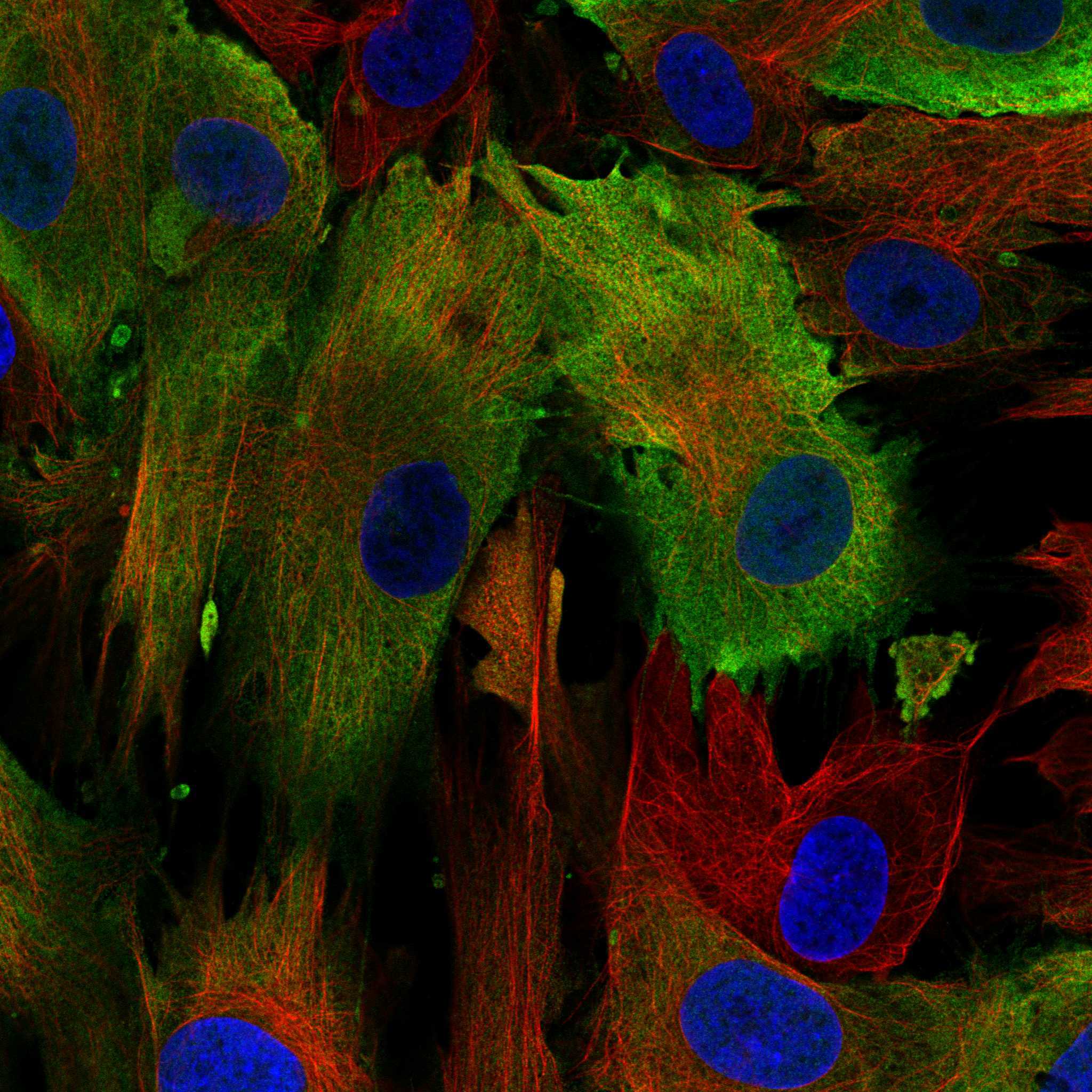
- Immunofluorescence staining of BJ cells using the anti-S100A4 monoclonal antibody, showing specific staining in the plasma membrane in green. Microtubule- and nuclear probes are visualized in red and blue, respectively (where available).
- Sample type
- HUMAN
Enhanced validation
Supportive validation
- Submitted by
- Atlas Antibodies (provider)
- Enhanced method
- Orthogonal validation
- Main image

- Experimental details
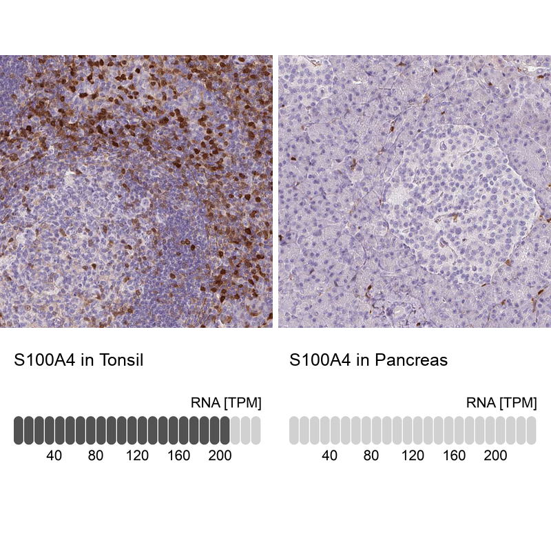
- Immunohistochemistry analysis in human tonsil and pancreas tissues using AMAb90599 antibody. Corresponding S100A4 RNA-seq data are presented for the same tissues.
- Sample type
- HUMAN
Supportive validation
- Submitted by
- Atlas Antibodies (provider)
- Main image

- Experimental details
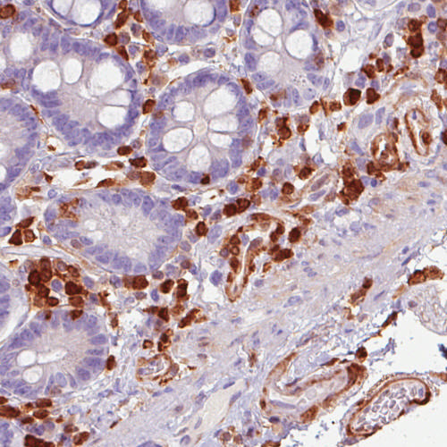
- Immunohistochemical staining of human duodenum shows immunoreactivity in endothelial and lymphoid cells.
- Submitted by
- Atlas Antibodies (provider)
- Main image
- Experimental details
- Immunohistochemical staining of human placenta shows immunoreactivity in decidual cells.

- Submitted by
- Atlas Antibodies (provider)
- Main image
- Experimental details
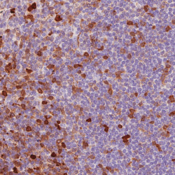
- Immunohistochemical staining of human tonsil shows moderate to strong immunoreactivity in a subset of lymphoid cells.
- Submitted by
- Atlas Antibodies (provider)
- Main image

- Experimental details
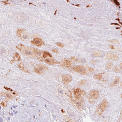
- Immunohistochemical staining of human prostate shows positivity in smooth muscle but not glandular cells.

- Submitted by
- Atlas Antibodies (provider)
- Main image
- Experimental details
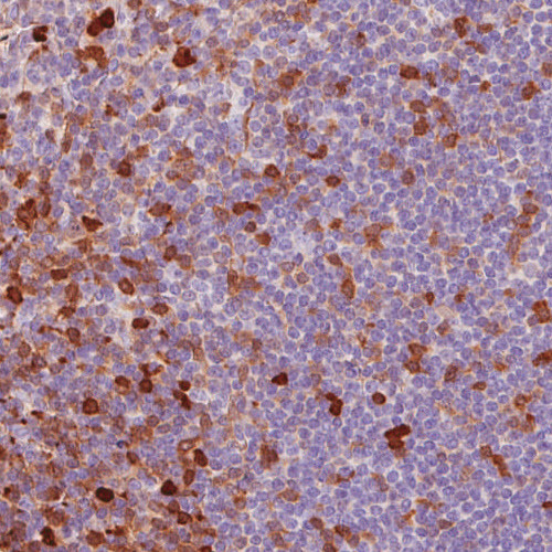
- Immunohistochemical staining of human tonsil shows moderate to strong cytoplasmic positivity, mainly in in non - germinal center cells.

- Submitted by
- Atlas Antibodies (provider)
- Main image
- Experimental details
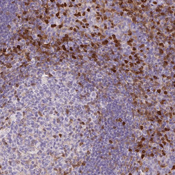
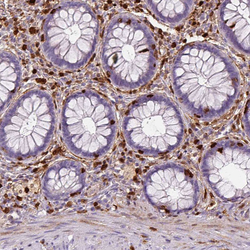
- Immunohistochemical staining of human rectum shows strong cytoplasmic positivity in lymphoid cells.

- Submitted by
- Atlas Antibodies (provider)
- Main image

- Experimental details
- Immunohistochemical staining of human endometrium shows strong cytoplasmic positivity in lymphoid cells.
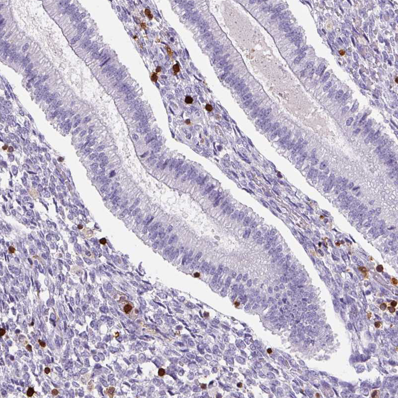
- Submitted by
- Atlas Antibodies (provider)
- Main image

- Experimental details
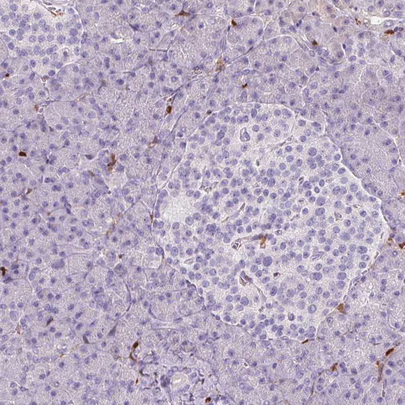
- Immunohistochemical staining of human pancreas shows no positivity in either Langerhans islets or exocrine glandular cells as expected.