Explore
 Validate
 Learn
Learn Western blot
 ELISA
ELISAAntibody data
- Antibody Data
- Antigen structure
- References [0]
- Comments [0]
- Validations
- Western blot [3]
- Immunocytochemistry [1]
Submit
Validation data
Reference
Comment
Report error
- Product number
- DM3507 - Provider product page

- Provider
- Acris Antibodies GmbH
- Proper citation
- Acris Antibodies GmbH Cat#DM3507, RRID:AB_1008464
- Product name
- anti VEGFR-1 / Flt-1
- Antibody type
- Monoclonal
- Antigen
- Recombinant Human soluble extracellular Flt-1 protein (D5) (Cat.-No DA3539)
- Reactivity
- Human, Mouse
- Host
- Mouse
- Isotype
- IgG
- Antibody clone number
- FLTEWI (EWI)
- Vial size
- 0.1 mg
No comments: Submit comment
Supportive validation
- Submitted by
- Acris Antibodies GmbH (provider)
- Main image
- Experimental details
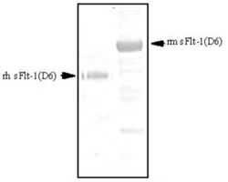
- Figure 1. Western Blot analysis showing cross reactivity of VEGFR-1/Flt-1 antibody Cat.-No DM3507 with recombinant Murine sFlt-1

- Submitted by
- Acris Antibodies GmbH (provider)
- Main image

- Experimental details
- Figure 1. Western Blot analysis showing cross reactivity of VEGFR-1/Flt-1 antibody Cat.-No DM3507 with recombinant Murine sFlt-1

- Submitted by
- Acris Antibodies GmbH (provider)
- Main image

- Experimental details
- Figure 2. Western Blot analysis showing cross reactivity of VEGFR-1/Flt-1 antibody Cat.-No DM3507 with recombinant Human endogenous sFlt-1 (esFlt-1) and sFlt-1(D5). There is no cross reactivity with recombinant Human sKDR.

Supportive validation
- Submitted by
- Acris Antibodies GmbH (provider)
- Main image
- Experimental details
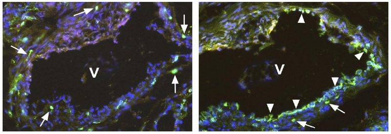
- Figure 3. Immunofluorescence staining (green) using Polyclonal antibody directed against the C-terminal end of native soluble VEGFR-1/Flt-1 Cat.-No DP3522 (Left panel) and Monoclonal antibody directed against the extracellular domain of the membrane-bound VEGFR-1/Flt-1 Cat.-No DM3507 (Right panel). You see two neighboring sections of a Human Vein (V), located near a hemangioma. The antibody against the soluble VEGFR-1/Flt-1 marked single cells (arrows) within the media and adventitia of the vein. The antibody against the membrane-bound VEGFR-1/Flt-1 marked single cells (arrows) and the endothelium (arrowhead) of the vein. Cell nuclei are stained with Dapi (blue). The experiment was performed by K. Butler and J. Wilting, University Göttingen, Germany.