Explore
 Validate
 Learn
Learn Western blot
 Immunoelectron microscopy
Antibody data
- Antibody Data
- Antigen structure
- References [0]
- Comments [0]
- Validations
- Western blot [2]
- Immunocytochemistry [1]
- Immunohistochemistry [2]
Submit
Validation data
Reference
Comment
Report error
- Product number
- PA5-111834 - Provider product page

- Provider
- Invitrogen Antibodies
- Product name
- GLP1R (extracellular) Polyclonal Antibody
- Antibody type
- Polyclonal
- Antigen
- Synthetic peptide
- Reactivity
- Human, Mouse, Rat
- Host
- Rabbit
- Isotype
- IgG
- Vial size
- 50 µL
- Concentration
- 0.8 mg/mL
- Storage
- -20°C
No comments: Submit comment
Supportive validation
- Submitted by
- Invitrogen Antibodies (provider)
- Main image

- Experimental details
- Western Blot analysis of GLP1R was performed in rat pancreas lysate (lanes 1 and 5), mouse preadipocyte 3T3-L1 lysate (lanes 2 and 6), rat pancreatic islet cell line RIN-5F lysate (lanes 3 and 7) and human pancreatic carcinoma PANC-1 lysate (lanes 4 and 8). Lane 1-4: GLP1R (extracellular) Antibody (Product # PA5-111834) at a dilution of 1:200. Lane 5-8: GLP1R (extracellular) Antibody preincubated with the negative control antigen.

Supportive validation
- Submitted by
- Invitrogen Antibodies (provider)
- Main image

- Experimental details
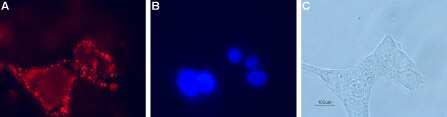
- Immunocytochemistry-Immunofluorescence analysis of GLP1R in live intact rat pancreatic islet cells. A) Cells were stained with GLP1R (extracellular) Antibody (Product # PA5-111834), (1:100), followed by goat anti-rabbit-AlexaFluor-594 secondary antibody (red). B) Cell nuclei were visualized with the membrane-permeable DNA dye Hoechst 33342 (blue staining). C) Live view of the cells.
Supportive validation
- Submitted by
- Invitrogen Antibodies (provider)
- Main image

- Experimental details
- Immunohistochemistry analysis of GLP1R in rat hypothalamus tissue sections using GLP1R (extracellular) Antibody (Product # PA5-111834) at a dilution of 1:200. Glucagon-like peptide 1 receptor (green) appears in the lateral hypothalamic region axonal profiles (vertical arrow) and nerve cell profiles (horizontal arrows). DAPI is used as the counterstain (blue).

- Submitted by
- Invitrogen Antibodies (provider)
- Main image

- Experimental details
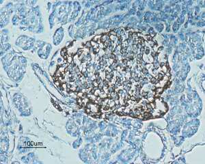
- Immunohistochemistry analysis of GLP1R in paraffin-embedded rat pancreas tissue sections using GLP1R (extracellular) Antibody (Product # PA5-111834) at a dilution of 1:100. Staining (brown color) is present in endocrine cells of the Isles of Langerhans. Hematoxilin is used as the counterstain.