 Explore
 Validate
 Learn
Learn Flow cytometry
 Other assay
Antibody data
- Antibody Data
- Antigen structure
- References [4]
- Comments [0]
- Validations
- Other assay [2]
Submit
Validation data
Reference
Comment
Report error
- Product number
- A15753 - Provider product page

- Provider
- Invitrogen Antibodies
- Product name
- CD81 Monoclonal Antibody (M38), FITC
- Antibody type
- Monoclonal
- Antigen
- Other
- Reactivity
- Human, Feline, Rabbit
- Host
- Mouse
- Conjugate
- Green dye
- Isotype
- IgG
- Antibody clone number
- M38
- Vial size
- 100 Tests
- Storage
- 4° C, store in dark
Submitted references Extracellular vesicles mediated exocytosis of antisense peptide nucleic acids.
Extracellular vesicle release from intestinal organoids is modulated by Apc mutation and other colorectal cancer progression factors.
CD19 Alterations Emerging after CD19-Directed Immunotherapy Cause Retention of the Misfolded Protein in the Endoplasmic Reticulum.
Evaluation and diagnostic potential of circulating extracellular vesicle-associated microRNAs in adrenocortical tumors.
Malik S, Saltzman WM, Bahal R
Molecular therapy. Nucleic acids 2021 Sep 3;25:302-315
Molecular therapy. Nucleic acids 2021 Sep 3;25:302-315
Extracellular vesicle release from intestinal organoids is modulated by Apc mutation and other colorectal cancer progression factors.
Szvicsek Z, Oszvald Á, Szabó L, Sándor GO, Kelemen A, Soós AÁ, Pálóczi K, Harsányi L, Tölgyes T, Dede K, Bursics A, Buzás EI, Zeöld A, Wiener Z
Cellular and molecular life sciences : CMLS 2019 Jun;76(12):2463-2476
Cellular and molecular life sciences : CMLS 2019 Jun;76(12):2463-2476
CD19 Alterations Emerging after CD19-Directed Immunotherapy Cause Retention of the Misfolded Protein in the Endoplasmic Reticulum.
Bagashev A, Sotillo E, Tang CH, Black KL, Perazzelli J, Seeholzer SH, Argon Y, Barrett DM, Grupp SA, Hu CC, Thomas-Tikhonenko A
Molecular and cellular biology 2018 Nov 1;38(21)
Molecular and cellular biology 2018 Nov 1;38(21)
Evaluation and diagnostic potential of circulating extracellular vesicle-associated microRNAs in adrenocortical tumors.
Perge P, Butz H, Pezzani R, Bancos I, Nagy Z, Pálóczi K, Nyírő G, Decmann Á, Pap E, Luconi M, Mannelli M, Buzás EI, Tóth M, Boscaro M, Patócs A, Igaz P
Scientific reports 2017 Jul 14;7(1):5474
Scientific reports 2017 Jul 14;7(1):5474
No comments: Submit comment
Supportive validation
- Submitted by
- Invitrogen Antibodies (provider)
- Main image
- Experimental details
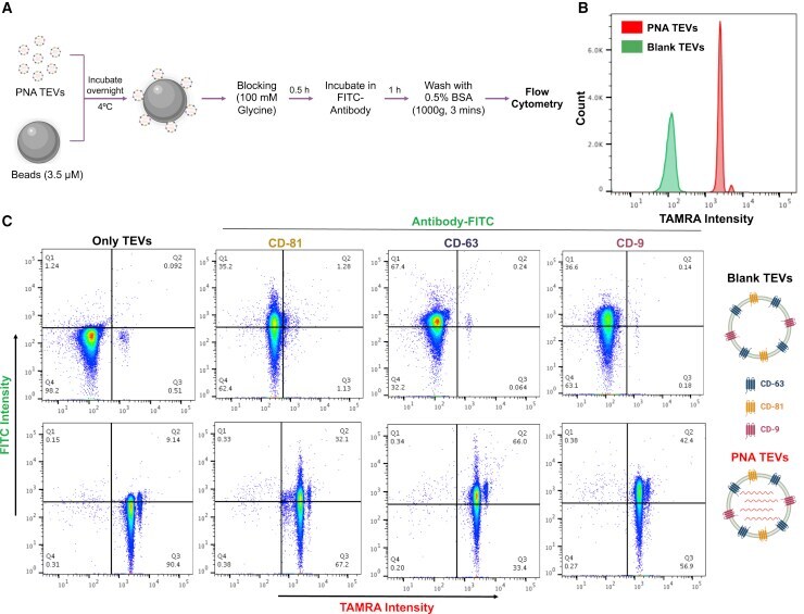
- Figure 3 Detection of TEVs via flow cytometry (A) Workflow depicting the steps used for detection of TEVs using flow cytometry. TEVs were collected from the supernatant of untreated HeLa cells (blank TEVs) or cells treated with PNA-155 (1 h) followed by washing and incubation in serum-free media for 24 h (PNA-TEVs). TEVs collected after ultracentrifugation were conjugated with latex/aldehyde beads followed by incubation of bead-TEVs complex with FITC-conjugated antibodies and detected by flow cytometry. (B) A representative histogram of blank TEVs and PNA TEVs. (C) Representative flow cytometry dot plots of blank TEVs and PNA TEVs showing staining with CD-63, CD-81, and CD-9 FITC-conjugated antibodies. First row represents blank TEVs and second row represents PNA TEVs. The 2 nd , 3 rd , and 4 th columns indicate staining with CD-81, CD-63, and CD-9 antibodies, respectively. 50,000 events were collected for each sample and all events were included in the dot plots.
- Conjugate
- Green dye
- Submitted by
- Invitrogen Antibodies (provider)
- Main image

- Experimental details
- FIG 8 CD19ex2 variants preferentially interact with ER/MHC-related proteins. (A) Immunoprecipitates of CD19 from lysates of transduced Nalm6 cell lines were subjected to mass spectrometry. The bars represent normalized total spectra for the identified protein targets. (B) Heat map showing proteins from the Reactome Adaptive Immune System gene set that exhibited differential binding to CD19, CD19-105R, and CD19-Deltaex2, as determined by mass spectrometry. (C) Double-stained live-cell flow cytometry using anti-CD19-PE and anti-CD81-FITC antibodies on Nalm6 parental and Nalm6-DeltaCD81 cell lines. (D) Immunoblotting of CD81, calnexin, calreticulin, UGCGL1, and PDI following immunoprecipitation with anti-CD19 antibody in lysates of the transduced cell lines. The CD19/CD81 double-KO Nalm6 cell line was used as a negative control. (E) Western blot for total levels of CD81, P97, and actin in lysates from the transduced cell lines. The CD19/CD81 double-KO Nalm6 cell line was used as a negative control. (F) Immunoblotting of CD81 following immunoprecipitation with anti-CD19 antibody in lysates from primary patient samples expressing CD19-WT (101 and 105) and CD19-105R immunotherapy-resistant isoforms.
- Conjugate
- Green dye
