Explore
 Validate
 Learn
Learn Western blot
Antibody data
- Antibody Data
- Antigen structure
- References [15]
- Comments [0]
- Validations
- Western blot [3]
- Immunocytochemistry [3]
- Immunohistochemistry [4]
- Other assay [4]
Submit
Validation data
Reference
Comment
Report error
- Product number
- MA1-940 - Provider product page

- Provider
- Invitrogen Antibodies
- Product name
- Galectin 3 Monoclonal Antibody (A3A12)
- Antibody type
- Monoclonal
- Antigen
- Recombinant full-length protein
- Description
- MA1-940 detects galectin-3 from human, mouse and rat tissues.
- Antibody clone number
- A3A12
- Concentration
- 1 mg/mL
Submitted references Impairment of the ER/mitochondria compartment in human cardiomyocytes with PLN p.Arg14del mutation.
Stimulation of β-adrenoceptors up-regulates cardiac expression of galectin-3 and BIM through the Hippo signalling pathway.
A novel phenylpyridazinone, T-3999, reduces the progression of autoimmune myocarditis to dilated cardiomyopathy.
Immunogold labelling in environmental scanning electron microscopy: applicative features for complementary cytological interpretation.
Partial inactivation of cardiac 14-3-3 protein in vivo elicits endoplasmic reticulum stress (ERS) and activates ERS-initiated apoptosis in ERS-induced mice.
Biofilm-like extracellular viral assemblies mediate HTLV-1 cell-to-cell transmission at virological synapses.
Shiga toxin 1 interaction with enterocytes causes apical protein mistargeting through the depletion of intracellular galectin-3.
Galectin-3 exerts cytokine-like regulatory actions through the JAK-STAT pathway.
Galectin-3 reduces the severity of pneumococcal pneumonia by augmenting neutrophil function.
Up-regulation of advanced glycated products receptors in the brain of diabetic rats is prevented by antioxidant treatment.
Galectin-3-positive cell infiltration in human diabetic nephropathy.
Galectin-3-positive cell infiltration in human diabetic nephropathy.
Galectin-3 messenger ribonucleic acid and protein are expressed in benign thyroid tumors.
Modulation of functional properties of galectin-3 by monoclonal antibodies binding to the non-lectin domains.
Expression of galectin-3 modulates T-cell growth and apoptosis.
Cuello F, Knaust AE, Saleem U, Loos M, Raabe J, Mosqueira D, Laufer S, Schweizer M, van der Kraak P, Flenner F, Ulmer BM, Braren I, Yin X, Theofilatos K, Ruiz-Orera J, Patone G, Klampe B, Schulze T, Piasecki A, Pinto Y, Vink A, Hübner N, Harding S, Mayr M, Denning C, Eschenhagen T, Hansen A
EMBO molecular medicine 2021 Jun 7;13(6):e13074
EMBO molecular medicine 2021 Jun 7;13(6):e13074
Stimulation of β-adrenoceptors up-regulates cardiac expression of galectin-3 and BIM through the Hippo signalling pathway.
Zhao WB, Lu Q, Nguyen MN, Su Y, Ziemann M, Wang LN, Kiriazis H, Puthalakath H, Sadoshima J, Hu HY, Du XJ
British journal of pharmacology 2019 Jul;176(14):2465-2481
British journal of pharmacology 2019 Jul;176(14):2465-2481
A novel phenylpyridazinone, T-3999, reduces the progression of autoimmune myocarditis to dilated cardiomyopathy.
Kamal FA, Watanabe K, Ma M, Abe Y, Elbarbary R, Kodama M, Aizawa Y
Heart and vessels 2011 Jan;26(1):81-90
Heart and vessels 2011 Jan;26(1):81-90
Immunogold labelling in environmental scanning electron microscopy: applicative features for complementary cytological interpretation.
Cafiero G, Papale F, Grimaldi A, Rosso F, Barbarisi M, Tortora C, Marino G, Barbarisi A
Journal of microscopy 2011 Jan;241(1):83-93
Journal of microscopy 2011 Jan;241(1):83-93
Partial inactivation of cardiac 14-3-3 protein in vivo elicits endoplasmic reticulum stress (ERS) and activates ERS-initiated apoptosis in ERS-induced mice.
Sari FR, Watanabe K, Widyantoro B, Thandavarayan RA, Harima M, Zhang S, Muslin AJ, Kodama M, Aizawa Y
Cellular physiology and biochemistry : international journal of experimental cellular physiology, biochemistry, and pharmacology 2010;26(2):167-78
Cellular physiology and biochemistry : international journal of experimental cellular physiology, biochemistry, and pharmacology 2010;26(2):167-78
Biofilm-like extracellular viral assemblies mediate HTLV-1 cell-to-cell transmission at virological synapses.
Pais-Correia AM, Sachse M, Guadagnini S, Robbiati V, Lasserre R, Gessain A, Gout O, Alcover A, Thoulouze MI
Nature medicine 2010 Jan;16(1):83-9
Nature medicine 2010 Jan;16(1):83-9
Shiga toxin 1 interaction with enterocytes causes apical protein mistargeting through the depletion of intracellular galectin-3.
Laiko M, Murtazina R, Malyukova I, Zhu C, Boedeker EC, Gutsal O, O'Malley R, Cole RN, Tarr PI, Murray KF, Kane A, Donowitz M, Kovbasnjuk O
Experimental cell research 2010 Feb 15;316(4):657-66
Experimental cell research 2010 Feb 15;316(4):657-66
Galectin-3 exerts cytokine-like regulatory actions through the JAK-STAT pathway.
Jeon SB, Yoon HJ, Chang CY, Koh HS, Jeon SH, Park EJ
Journal of immunology (Baltimore, Md. : 1950) 2010 Dec 1;185(11):7037-46
Journal of immunology (Baltimore, Md. : 1950) 2010 Dec 1;185(11):7037-46
Galectin-3 reduces the severity of pneumococcal pneumonia by augmenting neutrophil function.
Farnworth SL, Henderson NC, Mackinnon AC, Atkinson KM, Wilkinson T, Dhaliwal K, Hayashi K, Simpson AJ, Rossi AG, Haslett C, Sethi T
The American journal of pathology 2008 Feb;172(2):395-405
The American journal of pathology 2008 Feb;172(2):395-405
Up-regulation of advanced glycated products receptors in the brain of diabetic rats is prevented by antioxidant treatment.
Aragno M, Mastrocola R, Medana C, Restivo F, Catalano MG, Pons N, Danni O, Boccuzzi G
Endocrinology 2005 Dec;146(12):5561-7
Endocrinology 2005 Dec;146(12):5561-7
Galectin-3-positive cell infiltration in human diabetic nephropathy.
Kikuchi Y, Kobayashi S, Hemmi N, Ikee R, Hyodo N, Saigusa T, Namikoshi T, Yamada M, Suzuki S, Miura S
Nephrology, dialysis, transplantation : official publication of the European Dialysis and Transplant Association - European Renal Association 2004 Mar;19(3):602-7
Nephrology, dialysis, transplantation : official publication of the European Dialysis and Transplant Association - European Renal Association 2004 Mar;19(3):602-7
Galectin-3 messenger ribonucleic acid and protein are expressed in benign thyroid tumors.
Martins L, Matsuo SE, Ebina KN, Kulcsar MA, Friguglietti CU, Kimura ET
The Journal of clinical endocrinology and metabolism 2002 Oct;87(10):4806-10
The Journal of clinical endocrinology and metabolism 2002 Oct;87(10):4806-10
Modulation of functional properties of galectin-3 by monoclonal antibodies binding to the non-lectin domains.
Liu FT, Hsu DK, Zuberi RI, Hill PN, Shenhav A, Kuwabara I, Chen SS
Biochemistry 1996 May 14;35(19):6073-9
Biochemistry 1996 May 14;35(19):6073-9
Expression of galectin-3 modulates T-cell growth and apoptosis.
Yang RY, Hsu DK, Liu FT
Proceedings of the National Academy of Sciences of the United States of America 1996 Jun 25;93(13):6737-42
Proceedings of the National Academy of Sciences of the United States of America 1996 Jun 25;93(13):6737-42
No comments: Submit comment
Supportive validation
- Submitted by
- Invitrogen Antibodies (provider)
- Main image

- Experimental details
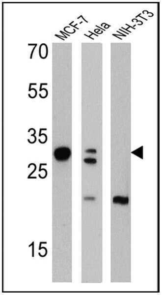
- Western blot analysis of Galectin 3 was performed by loading 25 µg of MCF-7 (Lane 1), Hela (Lane 2), and NIH-3T3 cell lysates (Lane 3) and a molecular weight protein ladder onto an SDS polyacrylamide gel. Proteins were transferred to a PVDF membrane and blocked with a blocking buffer at 4ºC overnight. The membrane was probed with a Galectin 3 monoclonal antibody (Product # MA1-940) at a dilution of 1:1000 overnight at 4°C, washed in TBST, and probed with an HRP-conjugated secondary antibody for 1 hr at room temperature in the dark. Chemiluminescent detection was performed using Pierce ECL Plus Western Blotting Substrate (Product # 32132). Results show a band at 30 kDa in MCF-7 and Hela cell lines.
- Submitted by
- Invitrogen Antibodies (provider)
- Main image
- Experimental details
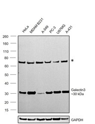
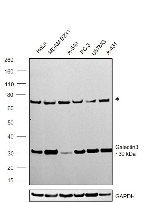
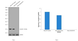
- Western blot was performed using Anti-Galectin 3 Monoclonal Antibody (A3A12)(Product # MA1-940) and a ~30kDa band corresponding to Galectin 3 was observed across cell lines tested . Whole cell extracts (30 µg lysate) of HeLa (Lane 1), MDA-MB-231 (Lane 2), A549 (Lane 3), PC-3 (Lane 4), U-87 MG (Lane 5), A-431 (Lane 6) were electrophoresed using NuPAGE™ 4-12% Bis-Tris Protein Gel (Product # NP0322BOX). Resolved proteins were then transferred onto a Nitrocellulose membrane (Product # LC2002) by iBlot® 2 Dry Blotting System (Product # IB21001). The blot was probed with the primary antibody (1:1000) and detected by chemiluminescence with Goat anti-Mouse IgG (H+L) Superclonal™ Recombinant Secondary Antibody, HRP (Product # A28177,1:4000) using the iBright FL 1000 (Product # A32752). Chemiluminescent detection was performed using Novex® ECL Chemiluminescent Substrate Reagent Kit (Product # WP20005).
- Submitted by
- Invitrogen Antibodies (provider)
- Main image
- Experimental details
- Knockout of LGALS3 was achieved by CRISPR-Cas9 genome editing using LentiArray™ Lentiviral sgRNA (Product # A32042, AssayID CRISPR855199_LV and CRISPR855204_LV) and LentiArray Cas9 Lentivirus (Product # A32064). Western blot analysis of LGALS3 was performed by loading 30 µg of HeLa wild type (Lane 1), HeLa CAS9 (Lane 2), HeLa LGALS3 KO (Lane 3) membrane extracts. The samples were electrophoresed using Novex® NuPAGE® 4-12% Bis-Tris Protein Gel (Product # NP0321BOX). Resolved proteins were then transferred onto a nitrocellulose membrane (Product # IB23001) by iBlot® 2 Dry Blotting System (Product # IB21001). The blot was probed with Anti-Galectin 3 Monoclonal Antibody (A3A12)(Product # MA1-940) using 1:1000 dilution and Goat anti-Mouse IgG (H+L), Superclonal™ Recombinant Secondary Antibody, HRP (Product # A28177, 1:4000 dilution) using the iBright FL 1000 (Product # A32752). Chemiluminescent detection was performed using Novex® ECL Chemiluminescent Substrate Reagent Kit (Product # WP20005). Loss of signal upon CRISPR mediated knockout (KO) using the LentiArray™ CRISPR product line confirms that antibody is specific to LGALS3.

Supportive validation
- Submitted by
- Invitrogen Antibodies (provider)
- Main image
- Experimental details
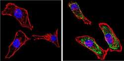
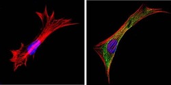
- Immunofluorescent analysis of Galectin 3 (green) showing staining in the cytoplasm and nucleus of HeLa cells. Formalin-fixed cells were permeabilized with 0.1% Triton X-100 in TBS for 5-10 minutes and blocked with 3% BSA-PBS for 30 minutes at room temperature. Cells were probed with a Galectin 3 monoclonal antibody (Product # MA1-940) in 3% BSA-PBS at a dilution of 1:100 and incubated overnight at 4 ºC in a humidified chamber. Cells were washed with PBST and incubated with a DyLight-conjugated secondary antibody in PBS at room temperature in the dark. F-actin (red) was stained with a fluorescent red phalloidin and nuclei (blue) were stained with Hoechst or DAPI. Images were taken at a magnification of 60x.
- Submitted by
- Invitrogen Antibodies (provider)
- Main image
- Experimental details
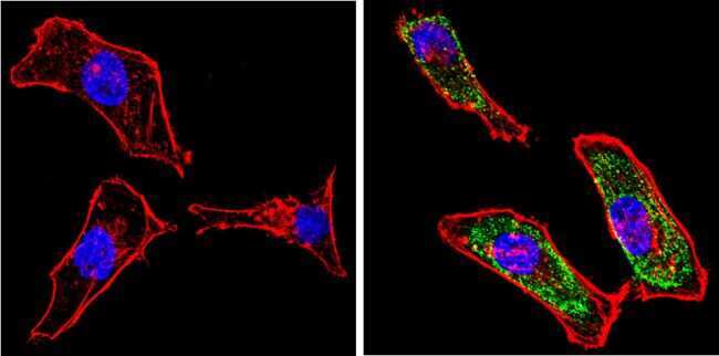
- Immunofluorescent analysis of Galectin 3 (green) showing staining in the cytoplasm and nucleus of MCF-7 cells. Formalin-fixed cells were permeabilized with 0.1% Triton X-100 in TBS for 5-10 minutes and blocked with 3% BSA-PBS for 30 minutes at room temperature. Cells were probed with a Galectin 3 monoclonal antibody (Product # MA1-940) in 3% BSA-PBS at a dilution of 1:100 and incubated overnight at 4 ºC in a humidified chamber. Cells were washed with PBST and incubated with a DyLight-conjugated secondary antibody in PBS at room temperature in the dark. F-actin (red) was stained with a fluorescent red phalloidin and nuclei (blue) were stained with Hoechst or DAPI. Images were taken at a magnification of 60x.

- Submitted by
- Invitrogen Antibodies (provider)
- Main image
- Experimental details
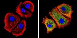
- Immunofluorescent analysis of Galectin 3 (green) showing staining in the cytoplasm and nucleus of NIH-3T3 cells. Formalin-fixed cells were permeabilized with 0.1% Triton X-100 in TBS for 5-10 minutes and blocked with 3% BSA-PBS for 30 minutes at room temperature. Cells were probed with a Galectin 3 monoclonal antibody (Product # MA1-940) in 3% BSA-PBS at a dilution of 1:100 and incubated overnight at 4 ºC in a humidified chamber. Cells were washed with PBST and incubated with a DyLight-conjugated secondary antibody in PBS at room temperature in the dark. F-actin (red) was stained with a fluorescent red phalloidin and nuclei (blue) were stained with Hoechst or DAPI. Images were taken at a magnification of 60x.

Supportive validation
- Submitted by
- Invitrogen Antibodies (provider)
- Main image
- Experimental details
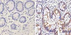
- Immunohistochemistry analysis of Galectin 3 showing positive staining in the nucleus and cytoplasm of paraffin-treated Human colon tissue (right) compared with a negative control in the absence of primary antibody (left). To expose target proteins, antigen retrieval method was performed using 10mM sodium citrate (pH 6.0) microwaved for 8-15 min. Following antigen retrieval, tissues were blocked in 3% H2O2-methanol for 15 min at room temperature, washed with ddH2O and PBS, and then probed with a Galectin 3 monoclonal antibody (Product # MA1-940) diluted by 3% BSA-PBS at a dilution of 1:100 overnight at 4°C in a humidified chamber. Tissues were washed extensively PBST and detection was performed using an HRP-conjugated secondary antibody followed by colorimetric detection using a DAB kit. Tissues were counterstained with hematoxylin and dehydrated with ethanol and xylene to prep for mounting.

- Submitted by
- Invitrogen Antibodies (provider)
- Main image
- Experimental details
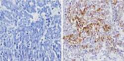
- Immunohistochemistry analysis of Galectin 3 showing positive staining in the nucleus and cytoplasm of paraffin-treated Human ovarian carcinoma (right) compared with a negative control in the absence of primary antibody (left). To expose target proteins, antigen retrieval method was performed using 10mM sodium citrate (pH 6.0) microwaved for 8-15 min. Following antigen retrieval, tissues were blocked in 3% H2O2-methanol for 15 min at room temperature, washed with ddH2O and PBS, and then probed with a Galectin 3 monoclonal antibody (Product # MA1-940) diluted by 3% BSA-PBS at a dilution of 1:100 overnight at 4°C in a humidified chamber. Tissues were washed extensively PBST and detection was performed using an HRP-conjugated secondary antibody followed by colorimetric detection using a DAB kit. Tissues were counterstained with hematoxylin and dehydrated with ethanol and xylene to prep for mounting.

- Submitted by
- Invitrogen Antibodies (provider)
- Main image

- Experimental details
- Immunohistochemistry analysis of Galectin 3 showing positive staining in the nucleus and cytoplasm of paraffin-treated Mouse colon tissue (right) compared with a negative control in the absence of primary antibody (left). To expose target proteins, antigen retrieval method was performed using 10mM sodium citrate (pH 6.0) microwaved for 8-15 min. Following antigen retrieval, tissues were blocked in 3% H2O2-methanol for 15 min at room temperature, washed with ddH2O and PBS, and then probed with a Galectin 3 monoclonal antibody (Product # MA1-940) diluted by 3% BSA-PBS at a dilution of 1:20 overnight at 4°C in a humidified chamber. Tissues were washed extensively PBST and detection was performed using an HRP-conjugated secondary antibody followed by colorimetric detection using a DAB kit. Tissues were counterstained with hematoxylin and dehydrated with ethanol and xylene to prep for mounting.

- Submitted by
- Invitrogen Antibodies (provider)
- Main image

- Experimental details
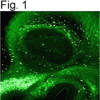
- Immunolocalization of galectin-3 in rat olfactory bulb using Product # MA1-940.
Supportive validation
- Submitted by
- Invitrogen Antibodies (provider)
- Main image

- Experimental details
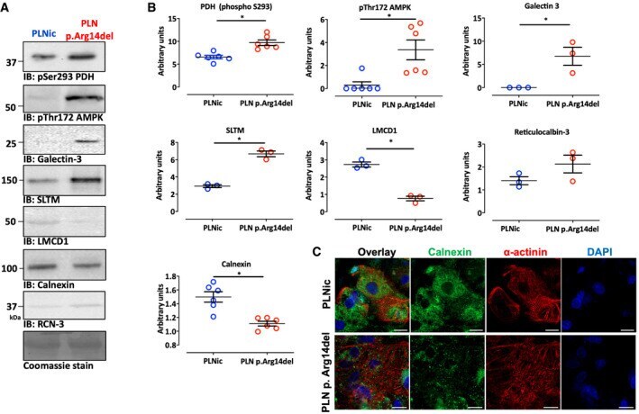
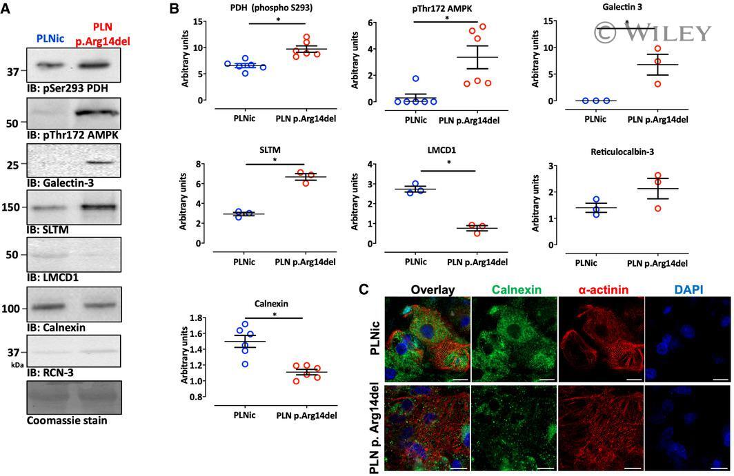
- EV1 Figure Investigation of protein expression and localization A, B Western immunoblots for phosphorylated pyruvate dehydrogenase at pS293 (PDH; n = 6, each replicate consists of a pool of 3-4 EHTs from 2 different separate batches), AMP-dependent protein kinase at Thr172 (AMPK; n = 6, each replicate consists of a pool of 3-4 EHTs from 2 different separate batches), galectin-3 ( n = 3; each replicate consists of a pool of 3-4 EHTs from 2 different separate batches), SAFB-like transcription modulator (SLTM; n = 3; each replicate consists of a pool of 3-4 EHTs from 2 different separate batches), LIM and cysteine-rich domains 1 (LMCD1; n = 3; each replicate consists of a pool of 3-4 EHTs from 2 different separate batches), calnexin ( n = 6; each replicate consists of a pool of 3-4 EHTs from 2 different separate batches), and reticulocalbin-3 (RCN-3; n = 3; each replicate consists of a pool of 3-4 EHTs from 2 different separate batches); Coomassie staining was used as a loading control, mean +- SEM, unpaired two-sided Student's t -test, * P < 0.05. C Immunofluorescence of 2D hiPSC-CM from PLNic and PLN p.Arg14del with antibodies recognizing calnexin (green), alpha-actinin (red), and DAPI staining for nuclei (blue); scale bar 20 um. Source data are available online for this figure.
- Submitted by
- Invitrogen Antibodies (provider)
- Main image
- Experimental details
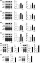
- Figure 5 PKA is involved in beta-adrenoceptor-induced YAP phosphorylation as well as Gal-3 and BIM up-regulation in H9c2 cells. H9c2 cells were pretreated with H89 (20 muM) or PKI 14-22 (10 muM) for 30 min prior to addition of isoprenaline (ISO; 3 muM) for 48 hr. Panels a and b are representative immunoblotting images and grouped data of p-YAP and YAP (a) or Gal-3 and BIM (b) in H9c2 cells after isoprenaline treatment alone or combined with H89. Panels c and d are representative immunoblotting images and grouped data of p-YAP and p-YAP/YAP ratio (c) or Gal-3 and BIM (d) in H9c2 cells after isoprenaline treatment alone or combined with PKI. For panels a-d, * P < .05, significantly different from CTL, # P < .05, significantly different from isoprenaline alone. H9c2 cells were treated with the AC activator forskolin (FSK, 10 muM) for 24 hr. Panels e and f show representative immunoblotting images and grouped data of p-YAP and YAP (e) or protein expression levels of Gal-3 and BIM (f). (g) Effect of isoprenaline (3 muM, 48 hr) or forskolin (10 muM, 24 hr) on Mst1 protein expression. For panels e-g, * P < .05, significantly different from CTL. For all data sets, n = 5 per group. Data shown are means +- SEM and were compared by two-tailed Student's t test (e-g) or one-way ANOVA (a-d) followed by Bonferroni post hoc test

- Submitted by
- Invitrogen Antibodies (provider)
- Main image

- Experimental details
- Figure 6 YAP is transcription co-activator and co-repressor, and its inhibition is associated with up-regulation of Gal-3 and BIM. Panels a-d show changes in cardiac gene expression by Mst1 overexpression. (a) Volcano plot of total RNA sequencing showing differentially regulated genes (DEGs) of Mst1-TG versus nTG hearts. (b) Volcano plot of YAP target genes according to ChIP-seq data. Activation of the Mst1(Hippo) signalling in the Mst1-TG heart leads to down- and up-regulated YAP target genes. For panels a and b, red points denote statistical significance (FDR < 0.05). (c) GSEA enrichment plot shows quantitatively that YAP target genes are collectively up-regulated in the Mst1-TG heart ( P < .0005). (d) List of top down- and up-regulated YAP-target genes in Mst1-TG hearts relative to nTG values. For panels a-d, n = 6 for nTG group and n = 7 for TG group. (e) H9c2 cells were transfected with YAP-siRNA or control-siRNA (1 muM for 72 hr). Representative immunoblotting images and changes in protein expression levels of YAP, BIM, and Gal-3. * P < .05, significantly different from CTL ( n = 6 per group). Data shown are means +- SEM and were compared by two-tailed Student's t test

- Submitted by
- Invitrogen Antibodies (provider)
- Main image

- Experimental details
- Figure EV1 Investigation of protein expression and localization A, B Western immunoblots for phosphorylated pyruvate dehydrogenase at pS293 (PDH; n = 6, each replicate consists of a pool of 3-4 EHTs from 2 different separate batches), AMP-dependent protein kinase at Thr172 (AMPK; n = 6, each replicate consists of a pool of 3-4 EHTs from 2 different separate batches), galectin-3 ( n = 3; each replicate consists of a pool of 3-4 EHTs from 2 different separate batches), SAFB-like transcription modulator (SLTM; n = 3; each replicate consists of a pool of 3-4 EHTs from 2 different separate batches), LIM and cysteine-rich domains 1 (LMCD1; n = 3; each replicate consists of a pool of 3-4 EHTs from 2 different separate batches), calnexin ( n = 6; each replicate consists of a pool of 3-4 EHTs from 2 different separate batches), and reticulocalbin-3 (RCN-3; n = 3; each replicate consists of a pool of 3-4 EHTs from 2 different separate batches); Coomassie staining was used as a loading control, mean +- SEM, unpaired two-sided Student's t -test, * P < 0.05. C Immunofluorescence of 2D hiPSC-CM from PLNic and PLN p.Arg14del with antibodies recognizing calnexin (green), alpha-actinin (red), and DAPI staining for nuclei (blue); scale bar 20 um. Source data are available online for this figure.