Explore
 Validate
 Learn
Learn Western blot
 Immunocytochemistry
Antibody data
- Antibody Data
- Antigen structure
- References [6]
- Comments [0]
- Validations
- Western blot [3]
- Immunohistochemistry [1]
- Flow cytometry [2]
Submit
Validation data
Reference
Comment
Report error
- Product number
- NB300-317 - Provider product page

- Provider
- Novus Biologicals
- Proper citation
- Novus Cat#NB300-317, RRID:AB_10002927
- Product name
- Rabbit Polyclonal xCT Antibody
- Antibody type
- Polyclonal
- Description
- Immunogen affinity purified.
- Reactivity
- Human, Mouse, Rat
- Host
- Rabbit
- Isotype
- IgG
- Vial size
- 0.1 ml
- Concentration
- 1 mg/ml
- Storage
- Store at 4C short term. Aliquot and store at -20C long term. Avoid freeze-thaw cycles.
Submitted references Dysregulation of Glutamate Transport Enhances Treg Function That Promotes VEGF Blockade Resistance in Glioblastoma.
RGS4 deficit in prefrontal cortex contributes to the behaviors related to schizophrenia via system x(c)(-)-mediated glutamatergic dysfunction in mice.
Rheostatic CD44 isoform expression and its association with oxidative stress in human malignant mesothelioma.
Signal transducer and activator of transcription 3 and 5 regulate system Xc- and redox balance in human breast cancer cells.
Increased expression of cystine/glutamate antiporter in multiple sclerosis.
Cystine-glutamate transporter SLC7A11 mediates resistance to geldanamycin but not to 17-(allylamino)-17-demethoxygeldanamycin.
Long Y, Tao H, Karachi A, Grippin AJ, Jin L, Chang YE, Zhang W, Dyson KA, Hou AY, Na M, Deleyrolle LP, Sayour EJ, Rahman M, Mitchell DA, Lin Z, Huang J
Cancer research 2020 Feb 1;80(3):499-509
Cancer research 2020 Feb 1;80(3):499-509
RGS4 deficit in prefrontal cortex contributes to the behaviors related to schizophrenia via system x(c)(-)-mediated glutamatergic dysfunction in mice.
Huang MW, Lin YJ, Chang CW, Lei FJ, Ho EP, Liu RS, Shyu WC, Hsieh CH
Theranostics 2018;8(17):4781-4794
Theranostics 2018;8(17):4781-4794
Rheostatic CD44 isoform expression and its association with oxidative stress in human malignant mesothelioma.
Chew SH, Okazaki Y, Akatsuka S, Wang S, Jiang L, Ohara Y, Ito F, Saya H, Sekido Y, Toyokuni S
Free radical biology & medicine 2017 May;106:91-99
Free radical biology & medicine 2017 May;106:91-99
Signal transducer and activator of transcription 3 and 5 regulate system Xc- and redox balance in human breast cancer cells.
Linher-Melville K, Haftchenary S, Gunning P, Singh G
Molecular and cellular biochemistry 2015 Jul;405(1-2):205-21
Molecular and cellular biochemistry 2015 Jul;405(1-2):205-21
Increased expression of cystine/glutamate antiporter in multiple sclerosis.
Pampliega O, Domercq M, Soria FN, Villoslada P, Rodríguez-Antigüedad A, Matute C
Journal of neuroinflammation 2011 Jun 3;8:63
Journal of neuroinflammation 2011 Jun 3;8:63
Cystine-glutamate transporter SLC7A11 mediates resistance to geldanamycin but not to 17-(allylamino)-17-demethoxygeldanamycin.
Liu R, Blower PE, Pham AN, Fang J, Dai Z, Wise C, Green B, Teitel CH, Ning B, Ling W, Lyn-Cook BD, Kadlubar FF, Sadée W, Huang Y
Molecular pharmacology 2007 Dec;72(6):1637-46
Molecular pharmacology 2007 Dec;72(6):1637-46
No comments: Submit comment
Supportive validation
- Submitted by
- Novus Biologicals (provider)
- Main image

- Experimental details
- Simple Western: xCT Antibody [NB300-317] - (1) ladder, (2) no lysate + xCT, 100ug/ml, (3) human brain frontal cortex membrane lysate,no primary antibody, (4-7) human brain lysates, 0.03mg/ml, xCT, 100ug/ml.

- Submitted by
- Novus Biologicals (provider)
- Main image
- Experimental details
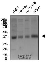
- Western Blot: xCT Antibody [NB300-317] - Total protein from human HeLa, Huvec, HCT-116 and A549 cells was separated on a 12% gel by SDS-PAGE, transferred to PVDF membrane and blocked in 5% non-fat milk in TBST. The membrane was probed with 2.0 ug/ml anti-xCT in block buffer and detected with an anti-rabbit HRP secondary antibody using chemiluminescence.

- Submitted by
- Novus Biologicals (provider)
- Main image

- Experimental details
- Western Blot: xCT Antibody [NB300-317] - Total protein from Human HeLa cells treated with and without 0.1 mM Diethyl Maleate for 24 hours was separated on a 12% gel by SDS-PAGE, transferred to PVDF membrane and blocked in 5% non-fat milk in TBST. The membrane was probed with 2.0 ug/ml anti-xCT in 1% non-fat milk in TBST and detected with an anti-rabbit HRP secondary antibody using chemiluminescence. Note the increase in xCT expression with treatment.

Supportive validation
- Submitted by
- Novus Biologicals (provider)
- Main image

- Experimental details
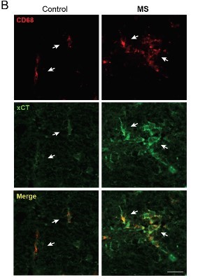
- Immunohistochemistry-Paraffin: xCT Antibody [NB300-317] - xCT expression is enhanced in CD68+ cells from MS spinal cord. CD68+ cells (arrows) show enhanced xCT expression in MS patients as compared to controls. CD68+ macrophages are round shaped and form clusters in MS patients, whereas in controls, CD68+ cells appear isolated and long shaped. Scale bar = 50 um. Image collected and cropped by CiteAb from the following publication (http://jneuroinflammation.biomedcentral.com/articles/10.1186/1742-2094-8-63), licensed under a CC-BY licence.
Supportive validation
- Submitted by
- Novus Biologicals (provider)
- Main image

- Experimental details
- Flow Cytometry: xCT Antibody [NB300-317] - An intracellular stain was performed on HeLa with NB300-317 and a matched isotype control. Cells were fixed with 4% PFA and then permeablized with 0.1% saponin. Cells were incubated in an antibody dilution of 2.5 ug/mL for 30 minutes at room temperature, followed by Rabbit IgG APC-conjugated Secondary Antibody (F0111, R&D Systems).

- Submitted by
- Novus Biologicals (provider)
- Main image

- Experimental details
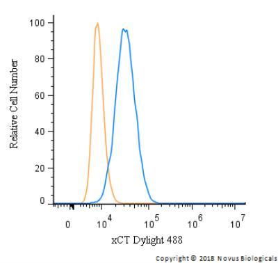
- Flow Cytometry: xCT Antibody [NB300-317] - An intracellular stain was performed on HeLa cells with NB300-317G (blue) and a matched isotype control (orange). Cells were fixed with 4% PFA and then permeabilized with 0.1% saponin. Cells were incubated in an antibody dilution of 5 ug/mL for 30 minutes at room temperature. Both antibodies were conjugated to DyLight 488.