 Explore
 Validate
 Learn
Learn Western blot
Antibody data
- Antibody Data
- Antigen structure
- References [0]
- Comments [0]
- Validations
- Western blot [4]
Submit
Validation data
Reference
Comment
Report error
- Product number
- MA5-27216 - Provider product page

- Provider
- Invitrogen Antibodies
- Product name
- SIRT1 Monoclonal Antibody (OTI1H1)
- Antibody type
- Monoclonal
- Antigen
- Recombinant protein fragment
- Reactivity
- Human, Mouse
- Host
- Mouse
- Isotype
- IgG
- Antibody clone number
- OTI1H1
- Vial size
- 100 µL
- Concentration
- 1 mg/mL
- Storage
- -20° C, Avoid Freeze/Thaw Cycles
No comments: Submit comment
Supportive validation
- Submitted by
- Invitrogen Antibodies (provider)
- Main image

- Experimental details
- Western blot analysis of SIRT1 in HEK293T cells in untransfected (Left lane) and transfected (Right lane) samples using 5 µg per lane. The samples were separated by SDS-PAGE and probed with SIRT1 (Product # MA5-27216) monoclonal antibody.

- Submitted by
- Invitrogen Antibodies (provider)
- Main image
- Experimental details
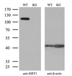
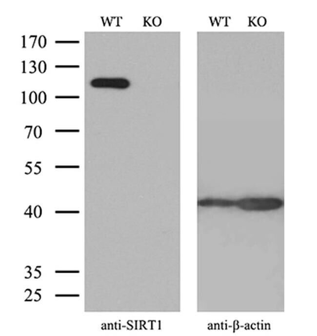
- Western blot analysis of SIRT1 in HeLa wild-type and knockout cells using 10 µg per lane. Samples were separated by SDS-PAGE and probed with SIRT1 (Product # MA5-27216) monoclonal antibody and beta-actin antibody with a dilution of 1:500 as a loading control.
- Submitted by
- Invitrogen Antibodies (provider)
- Main image

- Experimental details
- Western blot analysis of SIRT1 in HepG2, Jurkat, A549, HeLa cells using 35 µg per lane. Samples were probed with SIRT1 (Product # MA5-27216) monoclonal antibody at a dilution of 1:500.

- Submitted by
- Invitrogen Antibodies (provider)
- Main image

- Experimental details
- Western blot was performed using Anti-SIRT1 Monoclonal Antibody (OTI1H1) (Product # MA5-27216) and a 110 kDa band corresponding to SIRT1 was observed in all cell and tissue lysates tested. Modified whole cell extracts (1% SDS) (30 µg lysate) of MCF7 (Lane 1), HEK-293 (Lane 2), NTERA-2 cl.D1 (Lane 3) and Mouse Testis (Lane 4) were electrophoresed using Novex® NuPAGE® 4-12 % Bis-Tris gel (Product # NP0322BOX). Resolved proteins were then transferred onto a nitrocellulose membrane (Product # IB23001) by iBlot® 2 Dry Blotting System (Product # IB21001). The blot was probed with the primary antibody (1:500 dilution) and detected by chemiluminescence with Goat anti-Mouse IgG (H+L), Superclonal™ Recombinant Secondary Antibody, HRP (Product # A28177, 1:4000 dilution) using the iBright FL 1000 (Product # A32752). Chemiluminescent detection was performed using Novex® ECL Chemiluminescent Substrate Reagent Kit (Product # WP20005).
