Explore
 Validate
 Learn
Learn Western blot
 ELISA
ELISAAntibody data
- Antibody Data
- Antigen structure
- References [1]
- Comments [0]
- Validations
- Western blot [3]
- Immunocytochemistry [1]
- Other assay [3]
Submit
Validation data
Reference
Comment
Report error
- Product number
- 701155 - Provider product page

- Provider
- Invitrogen Antibodies
- Product name
- ANGPTL4 Recombinant Rabbit Monoclonal Antibody (1H12L19)
- Antibody type
- Monoclonal
- Antigen
- Other
- Description
- Recombinant rabbit monoclonal antibodies are produced using in vitro expression systems. The expression systems are developed by cloning in the specific antibody DNA sequences from immunoreactive rabbits. Then, individual clones are screened to select the best candidates for production. The advantages of using recombinant rabbit monoclonal antibodies include: better specificity and sensitivity, lot-to-lot consistency, animal origin-free formulations, and broader immunoreactivity to diverse targets due to larger rabbit immune repertoire.
- Reactivity
- Human, Mouse
- Host
- Rabbit
- Isotype
- IgG
- Antibody clone number
- 1H12L19
- Vial size
- 100 µg
- Concentration
- 0.5 mg/mL
- Storage
- Store at 4°C short term. For long term storage, store at -20°C, avoiding freeze/thaw cycles.
Submitted references Propranolol exhibits activity against hemangiomas independent of beta blockade.
Sasaki M, North PE, Elsey J, Bubley J, Rao S, Jung Y, Wu S, Zou MH, Pollack BP, Kumar J, Singh H, Arbiser JL
NPJ precision oncology 2019;3:27
NPJ precision oncology 2019;3:27
No comments: Submit comment
Supportive validation
- Submitted by
- Invitrogen Antibodies (provider)
- Main image

- Experimental details
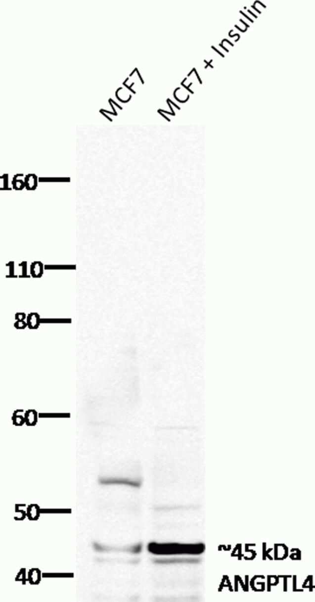
- Western blot analysis was performed on whole cell extracts (20 µg lysate) of MCF7 (lane 1) and MCF7 cells treated with insulin (lane 2) using the Xcell SureLock® Electrophoresis System and iBlot® Dry Blotting System. Endogenous ANGPTL4 at ~45 kDa was detected using Recombinant ANGPTL4 Rabbit Monoclonal Antibody (Product # 701155) at 0.5 µg/mL. Goat anti-Rabbit HRP Secondary Antibody (Product # G-21234) at 1:3000 dilution was used and the blot was developed using the chemiluminescence (ECL) technique.
- Submitted by
- Invitrogen Antibodies (provider)
- Main image
- Experimental details
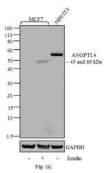
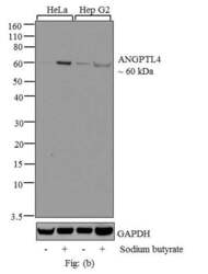
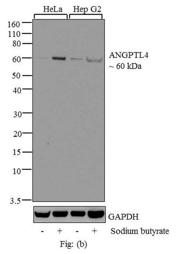
- Western blot analysis of ANGPTL4 was performed by loading 20 µg of MCF7 (lane1), MCF7 treated for 15 minutes with 100 µg/mL of Insulin (lane2) and NIH/3T3 (lane3) in Fig: (a), HeLa (lane1), HeLa treated for 12 hrs with 5 mM of sodium butyrate (lane2), Hep G2 (lane3) and Hep G2 treated for 12 hrs with 5 mM of sodium Buterate (lane4) in Fig: (b) cell lysate using NuPAGE® Novex® 10% Bis-Tris gel (Product # NP0301BOX), XCell SureLock Electrophoresis System (Product # EI0002), Novex® Sharp Pre-Stained Protein Standard (Product # LC5800), and iBlot® Dry Blotting System (Product # IB21001). Proteins were transferred to a nitrocellulose membrane and blocked with 5% skim milk for 1 hour at room temperature. ANGPTL4 was detected at ~45 and 60 kDa using ANGPTL4 Recombinant Rabbit Monoclonal Antibody (Product # 701155) at 2-3 µg/mL in 2.5% skim milk at 4°C overnight on a rocking platform. Goat anti-Rabbit IgG - HRP Secondary Antibody (Product # G-21234) at 1:5000 dilution was used and chemiluminescent detection was performed using Pierce™ ECL Western blotting Substrate (Product # 32106). The native full-length ANGPTL4 is a protein of 50 kDa. It has known splice variants of 45 kDa that are expressed in some cells, while in others ANGPTL4 is reported to be expressed as a 60 kDa glycosylated form of the native protein.

- Submitted by
- Invitrogen Antibodies (provider)
- Main image
- Experimental details
- Western blot analysis of ANGPTL4 was performed by loading 20 µg of MCF7 (lane1), MCF7 treated for 15 minutes with 100 µg/mL of Insulin (lane2) and NIH/3T3 (lane3) in Fig: (a), HeLa (lane1), HeLa treated for 12 hrs with 5 mM of sodium butyrate (lane2), Hep G2 (lane3) and Hep G2 treated for 12 hrs with 5 mM of sodium Buterate (lane4) in Fig: (b) cell lysate using NuPAGE® Novex® 10% Bis-Tris gel (Product # NP0301BOX), XCell SureLock Electrophoresis System (Product # EI0002), Novex® Sharp Pre-Stained Protein Standard (Product # LC5800), and iBlot® Dry Blotting System (Product # IB21001). Proteins were transferred to a nitrocellulose membrane and blocked with 5% skim milk for 1 hour at room temperature. ANGPTL4 was detected at ~45 and 60 kDa using ANGPTL4 Recombinant Rabbit Monoclonal Antibody (Product # 701155) at 2-3 µg/mL in 2.5% skim milk at 4°C overnight on a rocking platform. Goat anti-Rabbit IgG - HRP Secondary Antibody (Product # G-21234) at 1:5000 dilution was used and chemiluminescent detection was performed using Pierce™ ECL Western blotting Substrate (Product # 32106). The native full-length ANGPTL4 is a protein of 50 kDa. It has known splice variants of 45 kDa that are expressed in some cells, while in others ANGPTL4 is reported to be expressed as a 60 kDa glycosylated form of the native protein.
Supportive validation
- Submitted by
- Invitrogen Antibodies (provider)
- Main image
- Experimental details
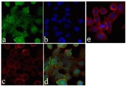
- Immunofluorescence analysis of ANGPTL4 was done on 70% confluent log phase HeLa cells. The cells were fixed with 4% paraformaldehyde for 15 minutes, permeabilized with 0.25% Triton X-100 for 10 minutes, and blocked with 5% BSA for 1 hour at room temperature. The cells were labeled with ANGPTL4 Recombinant Rabbit Monoclonal Antibody (Product # 701155) at 1 µg/mL in 1% BSA and incubated for 3 hours at room temperature and then labeled with Alexa Fluor 488 Goat anti-Rabbit IgG Secondary Antibody (Product # A-11008) at a dilution of 1:400 for 30 minutes at room temperature (Panel a: green). Nuclei (Panel b: blue) were stained with SlowFade® Gold Antifade Mountant DAPI (Product # S36938). F-actin (Panel c: red) was stained with Alexa Fluor 594 Phalloidin (Product # A12381). Panel d is a merged image showing membrane and cytoplasmic localization. Panel e shows no primary antibody control. The images were captured at 20X magnification.

Supportive validation
- Submitted by
- Invitrogen Antibodies (provider)
- Main image

- Experimental details
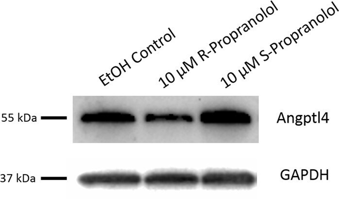
- Fig. 3 ANGPTL4 protein expression is reduced in R-propranolol-treated bEnd.3 cells. bEnd.3 cells were seeded 24 h prior to the R- or S-propranolol treatment. Culturing media were changed to complete media containing either 10 muM R- or S-propranolol or vehicle control of ethanol. After 24 h propranolol treatment, cells were subjected to western blotting and probed for ANGPTL4. R-propranolol differentially reduced the expression of ANGPTL4 as demonstrated by detection of the 55 kDa expected size bands. Loading control of GAPDH demonstrates that reduction is not due to the loading. Three independent experiments were performed, and a representative image is shown here. Other experimental results from western blotting is described in Supplementary Fig. 2 . Titration of R-propranolol ranging from 0 to 10 muM was performed to determine the optimal treatment concentration and described in Supplementary Fig. 3
- Submitted by
- Invitrogen Antibodies (provider)
- Main image
- Experimental details
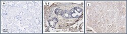
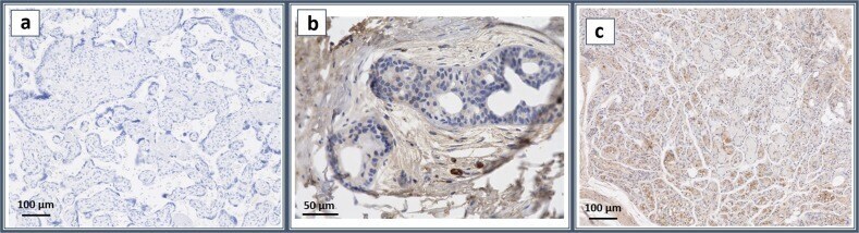
- Fig. 4 ANGPTL4 is expressed in hemangioma of infancy. Representative images of immunohistochemistry. The panels represent ( a ) no antibody control, ( b ) human infant foreskin sample stained with ANGPTL4, and ( c ) infantile hemangioma sample stained with Angptl4. The scale bars represent 100 mum ( a , c ) and 50 mum ( b ). Infantile hemangioma samples highly express ANGPTL4 proteins in endothelial cells, while human foreskin sample consists of largely negative cells surrounded by artifactually stained tissue
- Submitted by
- Invitrogen Antibodies (provider)
- Main image

- Experimental details
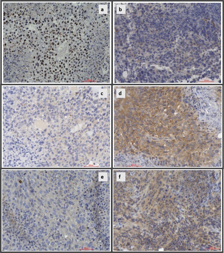
- Fig. 8 R-propranolol alters changes in the expression of proteins identified in RNAseq, validating the findings. Immunohistochemistry for ANGPTL4, BHMT, and APOA1 were performed on paraffin-embedded samples of R-propranolol- or ethanol vehicle-treated bEnd.3 murine tumor to validate the differential expression analysis results obtained using RNAseq. Nuclear expression of ANGPTL4 was markedly reduced in R-propranolol-treated animals while BHMT and APOA1 expression was increased in the experimental group, supporting the RNAseq findings. a , b Control- and R-propranolol-treated tumor samples stained with ANGPTL4. c , d Control- and R-propranolol-treated tumor samples stained with BHMT. e , f Control- and R-propranolol-treated tumor samples stained with APOA1. Scale bars indicate 50 mum in all panels