Explore
 Validate
 Learn
Learn Western blot
Antibody data
- Antibody Data
- Antigen structure
- References [0]
- Comments [0]
- Validations
- Western blot [5]
- Immunocytochemistry [2]
Submit
Validation data
Reference
Comment
Report error
- Product number
- 702060 - Provider product page

- Provider
- Invitrogen Antibodies
- Product name
- FASN Recombinant Rabbit Monoclonal Antibody (10H14L16)
- Antibody type
- Monoclonal
- Antigen
- Other
- Description
- This antibody is predicted to react with Monkey and galago.
- Antibody clone number
- 10H14L16
- Concentration
- 0.5 mg/mL
No comments: Submit comment
Supportive validation
- Submitted by
- Invitrogen Antibodies (provider)
- Main image

- Experimental details
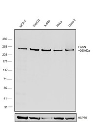
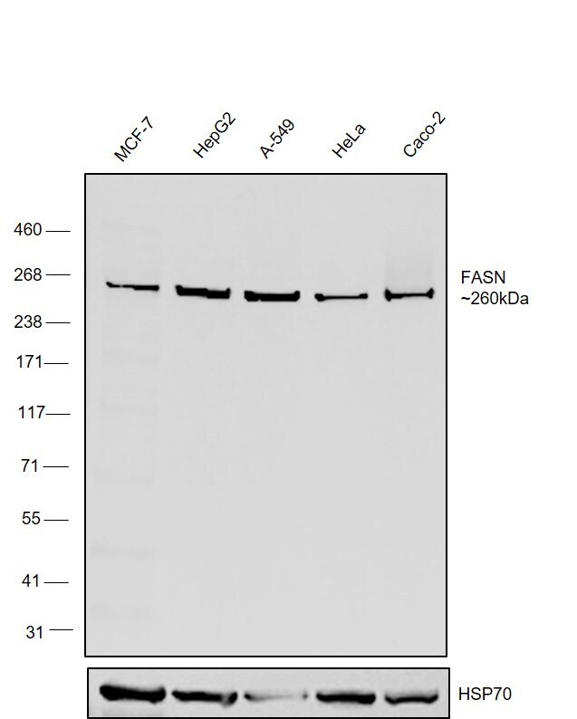
- Western blot was performed using Anti-FASN Recombinant Rabbit Monoclonal Antibody (10H14L16) (Product # 702060) and a ~260 kDa band corresponding to FASN was observed across cell lines tested . Whole cell extracts (30 µg lysate) of MCF7 (Lane 1), Hep G2 (Lane 2), A549 (Lane 3), HeLa (Lane 4), Caco-2 (Lane 5) were electrophoresed using NuPAGE™ 3-8% Tris-Acetate Protein Gel (Product # EA0378BOX). Resolved proteins were then transferred onto a Nitrocellulose membrane (Product # LC2001) by iBlot® 2 Dry Blotting System (Product # IB21001). The blot was probed with the primary antibody (1 µg/mL) and detected by chemiluminescence with Goat anti-Rabbit IgG (H+L) Superclonal™ Recombinant Secondary Antibody, HRP (Product # A27036,1:4000) using the iBright FL 1000 (Product # A32752). Chemiluminescent detection was performed using Novex® ECL Chemiluminescent Substrate Reagent Kit (Product # WP20005).

- Submitted by
- Invitrogen Antibodies (provider)
- Main image

- Experimental details
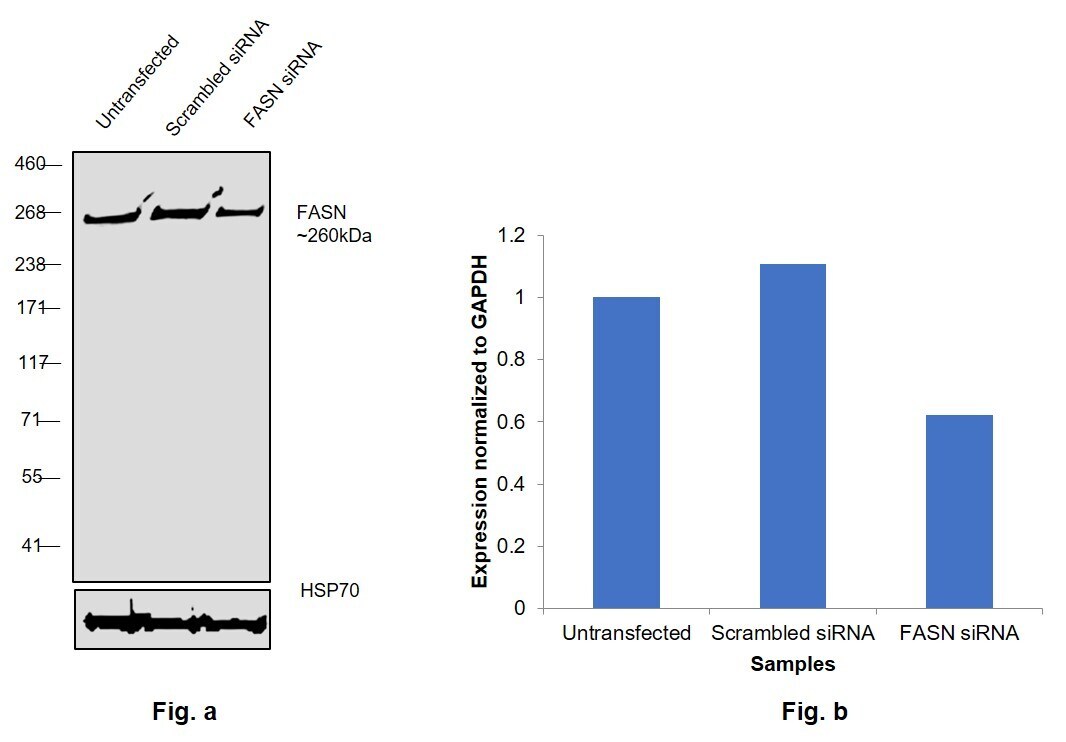
- Knockdown of FASN was achieved by transfecting Hep G2 with FASN specific siRNAs (Silencer® select Product # s5030. s5032). Western blot analysis (Fig. a) was performed using Whole cell extracts from the FASN knockdown cells (lane 3), non-targeting scrambled siRNA transfected cells (lane 2) and untransfected cells (lane 1). The blot was probed with FASN Recombinant Rabbit Monoclonal Antibody (10H14L16) (Product # 702060, 1:1000 ) and Goat anti-Rabbit IgG (H+L) Superclonal™ Recombinant Secondary Antibody, HRP (Product # A27036, 1:4000). Densitometric analysis of this western blot is shown in histogram (Fig. b). Decrease in signal upon siRNA mediated knock down confirms that antibody is specific to FASN.
- Submitted by
- Invitrogen Antibodies (provider)
- Main image
- Experimental details
- CRISPR-Cas9 mediated genome editing ofFASN (as confirmed by next generation sequencing) was achieved by using LentiArray™ Lentiviral sgRNA (Product # A32042, Assay ID CRISPR806223_LV) and LentiArray Cas9 Lentivirus (Product # A32064). Fig (a) Western blot analysis of FASN was performed by loading 30 µg of HepG2 Wild Type (Lane 1), HepG2 Cas9 (Lane 2) and HepG2 Cas9 cells transduced with FASN Lentiviral sgRNA (Lane 3) whole cell extracts. The samples were electrophoresed using NuPAGE™ 3-8% Tris-Acetate Protein Gel (Product # EA0378BOX). Resolved proteins were then transferred onto a nitrocellulose membrane (Product # IB23001) by iBlot® 2 Dry Blotting System (Product # IB21001). The blot was probed with Anti-FASN Recombinant Rabbit Monoclonal Antibody (10H14L16) (Product # 702060) using 1:1000 dilution and Goat anti-Rabbit IgG (H+L) Superclonal™ Recombinant Secondary Antibody, HRP (Product # A27036 1:5000 dilution).Chemiluminescent detection was performed using Novex® ECL Chemiluminescent Substrate Reagent Kit (Product # WP20005). A loss of signal in sgRNA transduced cells using the LentiArray™ CRISPR product line confirms that antibody is specific toFASN (Fig (b)).

- Submitted by
- Invitrogen Antibodies (provider)
- Main image
- Experimental details
- Western blot was performed using Anti-FASN Recombinant Rabbit Monoclonal Antibody (10H14L16) (Product # 702060) and a ~260 kDa band corresponding to FASN was observed across cell lines tested . Whole cell extracts (30 µg lysate) of MCF7 (Lane 1), Hep G2 (Lane 2), A549 (Lane 3), HeLa (Lane 4), Caco-2 (Lane 5) were electrophoresed using NuPAGE™ 3-8% Tris-Acetate Protein Gel (Product # EA0378BOX). Resolved proteins were then transferred onto a Nitrocellulose membrane (Product # LC2001) by iBlot® 2 Dry Blotting System (Product # IB21001). The blot was probed with the primary antibody (1 µg/mL) and detected by chemiluminescence with Goat anti-Rabbit IgG (H+L) Superclonal™ Recombinant Secondary Antibody, HRP (Product # A27036,1:4000) using the iBright FL 1000 (Product # A32752). Chemiluminescent detection was performed using Novex® ECL Chemiluminescent Substrate Reagent Kit (Product # WP20005).
- Submitted by
- Invitrogen Antibodies (provider)
- Main image

- Experimental details
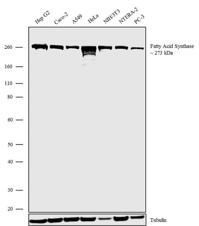
- Western blot analysis was performed on Membrane enriched extracts (30 µg lysate) of Hep G2 (Lane 1), Caco-2 (Lane 2), A549 (Lane 3), HeLa (Lane 4), NIH/3T3 (Lane 5), NTERA-2 (Lane 6) and PC-3 (Lane 7). The blots were probed with Anti-Fatty Acid Synthase Recombinant Rabbit Monoclonal Antibody (Product # 702060, 1-2 µg/mL) and detected by chemiluminescence using Goat anti-Rabbit IgG (H+L) Superclonal Secondary Antibody, HRP conjugate (Product # A27036, 0.4 µg/mL, 1:2500 dilution). A 273 kDa band corresponding to Fatty Acid Synthase was observed across the cell ines tested. Known quantity of protein samples were electrophoresed using Novex®NuPAGE®4-12% Bis-Tris gel (Product # NP0321BOX), XCell SureLock Electrophoresis System (Product # EI0002) and Novex® Sharp Pre-Stained Protein Standard (Product # LC5800). Resolved proteins were then transferred onto a nitrocellulose membrane with wet transfer method. The membrane was probed with the relevant primary and secondary Antibody following blocking with 5% skimmed milk. Chemiluminescent detection was performed using Pierce™ ECL Western blotting Substrate (Product # 32106).
Supportive validation
- Submitted by
- Invitrogen Antibodies (provider)
- Main image
- Experimental details
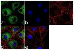
- For immunofluorescence analysis, Hep G2 cells were fixed and permeabilized for detection of endogenous Fatty acid synthase using Fatty Acid Synthase Recombinant Rabbit Monoclonal Antibody (Product # 702060, 2 µg/mL) and labeled with Goat anti-Rabbit IgG (H+L) Superclonal Secondary Antibody, Alexa Fluor® 488 conjugate (Product # A27034, 1:2000). Panel a) shows representative cells that were stained for detection and localization of Fatty acid synthase protein (green), Panel b) is stained for nuclei (blue) using SlowFade® Gold Antifade Mountant with DAPI (Product # S36938). Panel c) represents cytoskeletal F-actin staining using Alexa Fluor® 555 Rhodamine Phalloidin (Product # R415, 1:300). Panel d) is a composite image of Panels a, b and c clearly demonstrating cytoplasmic localization of Fatty acid synthase. Panel e) represents control cells with no primary antibody to assess background. The images were captured at 60X magnification.
- Submitted by
- Invitrogen Antibodies (provider)
- Main image
- Experimental details
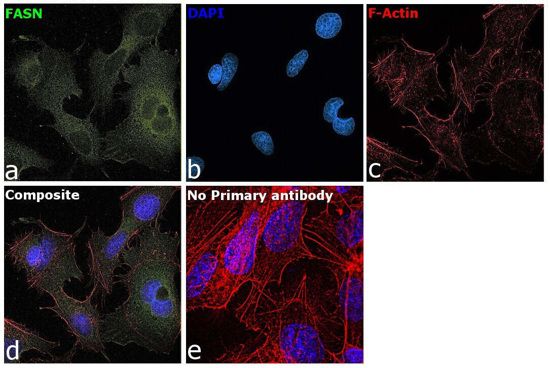
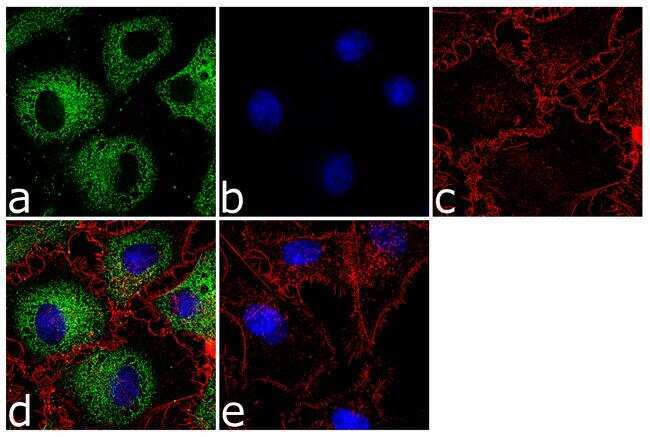
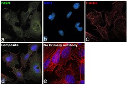
- Immunofluorescence analysis of FASN was performed using 70% confluent log phase Hep G2 cells. The cells were fixed with 4% paraformaldehyde for 10 minutes, permeabilized with 0.1% Triton™ X-100 for 15 minutes, and blocked with 2% BSA for 45 minutes at room temperature. The cells were labeled with FASN Recombinant Rabbit Monoclonal Antibody (10H14L16) (Product # 702060) at 2 µg/mL in 0.1% BSA, incubated at 4 degree celsius overnight and then labeled with Donkey anti-Rabbit IgG (H+L) Highly Cross-Adsorbed Secondary Antibody, Alexa Fluor Plus 488 (Product # A32790), (1:2000), for 45 minutes at room temperature (Panel a: Green). Nuclei (Panel b:Blue) were stained with ProLong™ Diamond Antifade Mountant with DAPI (Product # P36962). F-actin (Panel c: Red) was stained with Rhodamine Phalloidin (Product # R415, 1:300). Panel d represents the merged image showing cytoplasmic localization. Panel e represents control cells with no primary antibody to assess background. The images were captured at 60X magnification.