 Explore
 Validate
 Learn
Learn Western blot
Antibody data
- Antibody Data
- Antigen structure
- References [0]
- Comments [0]
- Validations
- Western blot [6]
- Immunocytochemistry [4]
- Immunohistochemistry [3]
- Other assay [1]
Submit
Validation data
Reference
Comment
Report error
- Product number
- PA5-22111 - Provider product page

- Provider
- Invitrogen Antibodies
- Product name
- FASN Polyclonal Antibody
- Antibody type
- Polyclonal
- Antigen
- Recombinant protein fragment
- Description
- Recommended positive controls: 293T, A431, HeLa, HepG2, Molt-4, Mouse liver.
- Concentration
- 1 mg/mL
No comments: Submit comment
Supportive validation
- Submitted by
- Invitrogen Antibodies (provider)
- Main image
- Experimental details
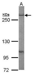
- Western blot analysis of Fatty Acid Synthase using 50 µg of mouse liver lysate. Samples were loaded onto a 5% SDS-PAGE gel and probed with a Fatty Acid Synthase polyclonal antibody (Product # PA5-22111) at a dilution of 1:1000.

- Submitted by
- Invitrogen Antibodies (provider)
- Main image

- Experimental details
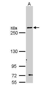
- Western Blot using FASN Polyclonal Antibody (Product # PA5-22111). Sample (30 µg of whole cell lysate). Lane A: Molt-4 .5% SDS PAGE. FASN Polyclonal Antibody (Product # PA5-22111) diluted at 1:1,000.
- Submitted by
- Invitrogen Antibodies (provider)
- Main image

- Experimental details
- Western Blot using FASN Polyclonal Antibody (Product # PA5-22111). Unstimulatd and stimulatd 3T3-L1 whole cell extracts (20 µg) were separated by 5% SDS-PAGE, and the membrane was blotted with FASN Polyclonal Antibody (Product # PA5-22111) diluted at 1:500. The HRP-conjugated anti-rabbit IgG antibody was used to detect the primary antibody. (The differentiation stimulated medium is composed by basal medium, 10% FBS, 50 µg/mL gentamicin, 1 nM L-glutamin, 500 µM IBMX, 1 µM dexamethasone, 2 µM rosiglitazone and 1 µg/mL insulin.).

- Submitted by
- Invitrogen Antibodies (provider)
- Main image
- Experimental details
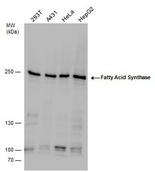
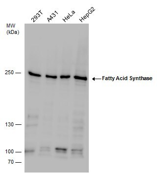
- FASN Polyclonal Antibody detects Fatty Acid Synthase protein by western blot analysis. Various whole cell extracts (30 µg) were separated by 5% SDS-PAGE, and the membrane was blotted with FASN Polyclonal Antibody (Product # PA5-22111) diluted by 1:1,000.
- Submitted by
- Invitrogen Antibodies (provider)
- Main image
- Experimental details
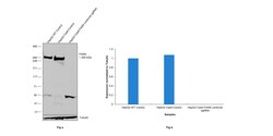
- CRISPR-Cas9 mediated genome editing ofFASN (as confirmed by next generation sequencing) was achieved by using LentiArray™ Lentiviral sgRNA (Product # A32042, Assay ID CRISPR806223_LV) and LentiArray Cas9 Lentivirus (Product # A32064). Fig (a) Western blot analysis of FASN was performed by loading 30 µg of HepG2 wild type (Lane 1), HepG2 Cas9 (Lane 2) and HepG2 Cas9 cells transduced with FASN Lentiviral sgRNA (Lane 3) whole cell extracts. The samples were electrophoresed using NuPAGE™ 3-8% Tris-Acetate Protein Gel (Product # EA0378BOX). Resolved proteins were then transferred onto a nitrocellulose membrane (Product # IB23001) by iBlot® 2 Dry Blotting System (Product # IB21001). The blot was probed with Anti-FASN Polyclonal Antibody (Product # PA5-22111) using 1:1000 dilution and Goat anti-Rabbit IgG (H+L) Superclonal™ Recombinant Secondary Antibody, HRP (Product # A27036 1:5000 dilution).Chemiluminescent detection was performed using Novex® ECL Chemiluminescent Substrate Reagent Kit (Product # WP20005). A loss of signal in sgRNA transduced cells using the LentiArray™ CRISPR product line confirms that antibody is specific toFASN (Fig (b)). Uncharacterized band was observed in all the samples at ~90 kDa.
- Submitted by
- Invitrogen Antibodies (provider)
- Main image
- Experimental details
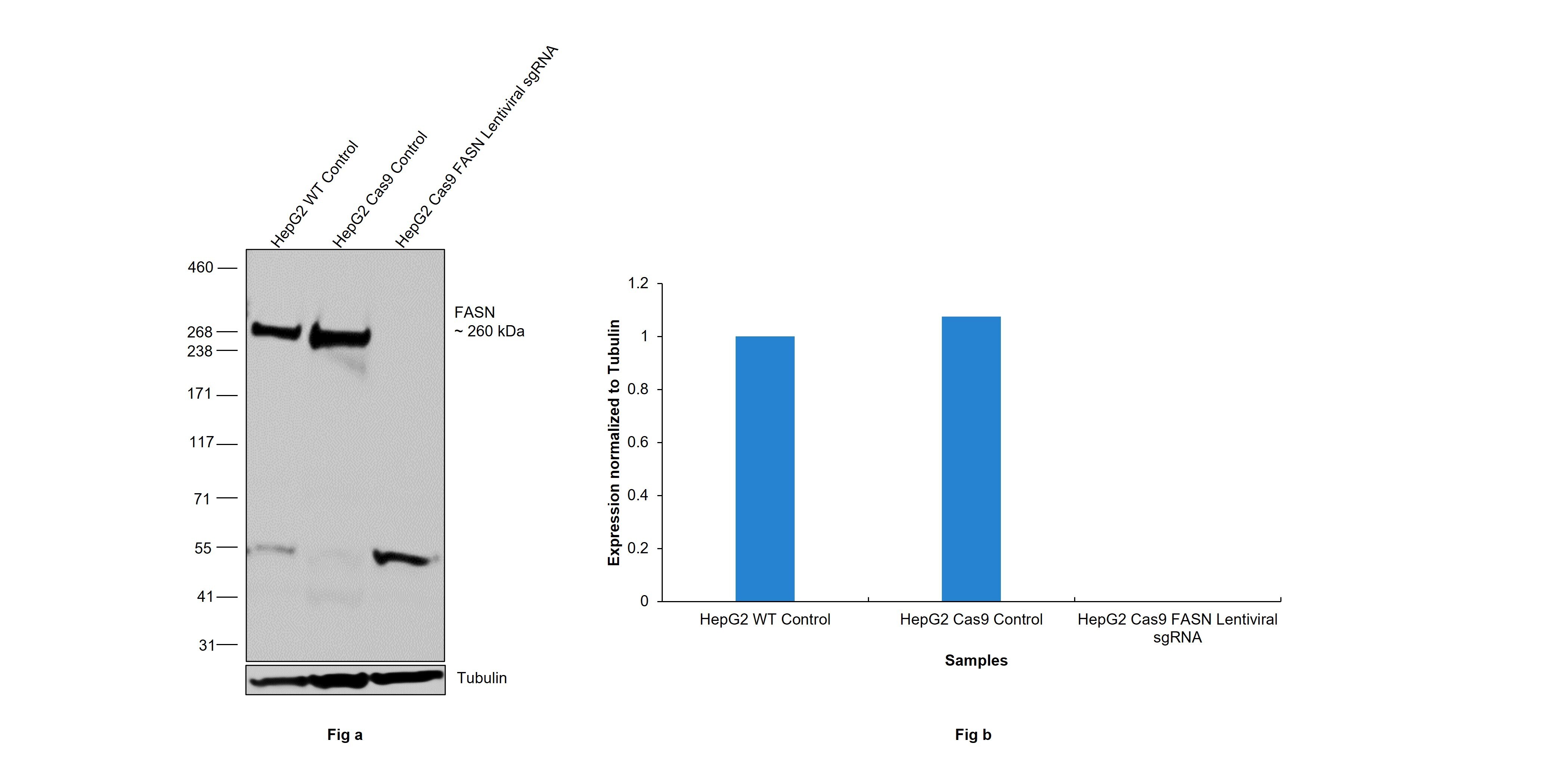
- Western blot was performed using Anti-FASN Polyclonal Antibody(Product # PA5-22111) and a ~260kDa band corresponding to FASN was observed across cell lines tested . Whole cell extracts (30 µg lysate) of MCF7 (Lane 1), Hep G2 (Lane 2), A549 (Lane 3), HeLa (Lane 4), Caco-2 (Lane 5), NTERA-2 cl.D1 (Lane 6), PC-3 (Lane 7) were electrophoresed using NuPAGE™ 3-8% Tris-Acetate Protein Gel (Product # EA0378BOX). Resolved proteins were then transferred onto a Nitrocellulose membrane (Product # LC2001) by iBlot® 2 Dry Blotting System (Product # IB21001). The blot was probed with the primary antibody (1:1000) and detected by chemiluminescence with Goat anti-Rabbit IgG (H+L) Superclonal™ Recombinant Secondary Antibody, HRP (Product # A27036,1:4000) using the iBright FL 1000 (Product # A32752). Chemiluminescent detection was performed using Novex® ECL Chemiluminescent Substrate Reagent Kit (Product # WP20005).

Supportive validation
- Submitted by
- Invitrogen Antibodies (provider)
- Main image
- Experimental details
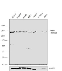
- Immunofluorescent analysis of Fatty Acid Synthase in paraformaldehyde-fixed HeLa cells using a Fatty Acid Synthase polyclonal antibody (Product # PA5-22111) (Green) at a 1:500 dilution. Alpha-tubulin filaments were labeled with Product # PA5-29281 (Red) at a 1:2000.

- Submitted by
- Invitrogen Antibodies (provider)
- Main image

- Experimental details
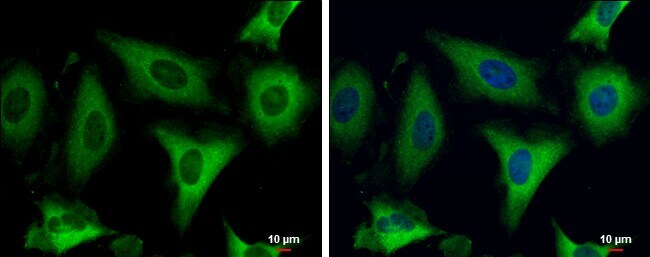
- Immunocytochemistry-Immunofluorescence analysis of FASN was performed in HeLa cells fixed in 4% paraformaldehyde at RT for 15 min. Green: FASN Polyclonal Antibody (Product # PA5-22111) diluted at 1:500. Blue: Hoechst 33342 staining. Scale bar = 10 µm.
- Submitted by
- Invitrogen Antibodies (provider)
- Main image
- Experimental details
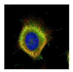
- Confocal immunofluorescence analysis (Olympus FV10i) of paraformaldehyde-fixed HeLa, using Fatty Acid Synthase antibody (Product # PA5-22111) (Green) at 1:500 dilution. Alpha-tubulin filaments were labeled with (Product # MA1-25054) (Red) at 1:2,000.

- Submitted by
- Invitrogen Antibodies (provider)
- Main image
- Experimental details
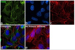
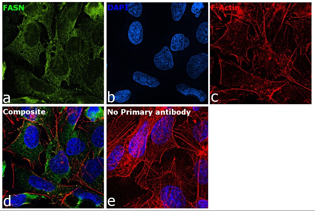
- Immunofluorescence analysis of FASN was performed using 70% confluent log phase Hep G2 cells. The cells were fixed with 4% paraformaldehyde for 10 minutes, permeabilized with 0.1% Triton™ X-100 for 15 minutes, and blocked with 2% BSA for 45 minutes at room temperature. The cells were labeled with FASN Polyclonal Antibody (Product # PA5-22111) at 1:200 in 0.1% BSA, incubated at 4 degree celsius overnight and then labeled with Donkey anti-Rabbit IgG (H+L) Highly Cross-Adsorbed Secondary Antibody, Alexa Fluor Plus 488 (Product # A32790), (1:2000), for 45 minutes at room temperature (Panel a: Blue). Nuclei (Panel b:Green) were stained with ProLong™ Diamond Antifade Mountant with DAPI (Product # P36962). F-actin (Panel c: Red) was stained with Rhodamine Phalloidin (Product # R415, 1:300). Panel d represents the merged image showing cytoplasmic localization. Panel e represents control cells with no primary antibody to assess background. The images were captured at 60X magnification.
Supportive validation
- Submitted by
- Invitrogen Antibodies (provider)
- Main image

- Experimental details
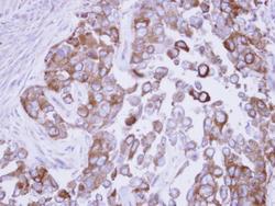
- FASN Polyclonal Antibody detects Fatty Acid Synthase protein at cytoplasm by immunohistochemical analysis. Sample: Paraffin-embedded mouse adrenal gland. Fatty Acid Synthase stained by FASN Polyclonal Antibody (Product # PA5-22111) diluted at 1:500. Antigen Retrieval: Citrate buffer, pH 6.0, 15 min.
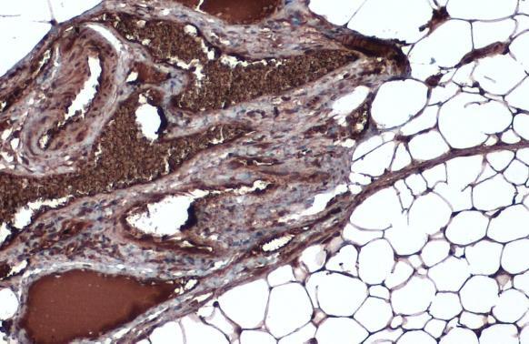
- Submitted by
- Invitrogen Antibodies (provider)
- Main image

- Experimental details
- FASN Polyclonal Antibody detects Fatty Acid Synthase protein at cytoplasm by immunohistochemical analysis. Sample: Paraffin-embedded mouse adrenal gland. Fatty Acid Synthase stained by FASN Polyclonal Antibody (Product # PA5-22111) diluted at 1:500. Antigen Retrieval: Citrate buffer, pH 6.0, 15 min.
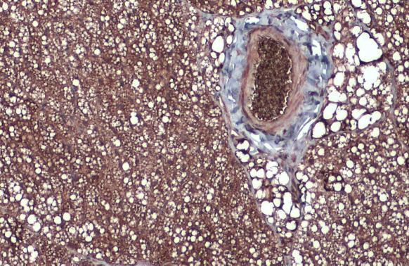
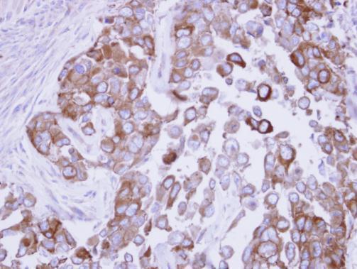
- Submitted by
- Invitrogen Antibodies (provider)
- Main image
- Experimental details
- Immunohistochemical analysis of paraffin-embedded OECM1 xenograft, using Fatty Acid Synthase (Product # PA5-22111) antibody at 1:500 dilution. Antigen Retrieval: EDTA based buffer, pH 8.0, 15 min.
Supportive validation
- Submitted by
- Invitrogen Antibodies (provider)
- Main image
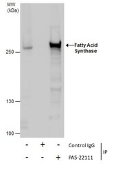
- Experimental details
- Immunoprecipitation of Fatty Acid Synthase was performed in HeLa whole cell extracts using 5 µg of FASN Polyclonal Antibody (Product # PA5-22111). Samples were transferred to a membrane and probed with FASN Polyclonal Antibody as a primary antibody and an HRP-conjugated anti-Rabbit IgG was used as a secondary antibody.
