 Explore
 Validate
 Learn
Learn Western blot
Antibody data
- Antibody Data
- Antigen structure
- References [0]
- Comments [0]
- Validations
- Western blot [1]
- Immunohistochemistry [2]
Submit
Validation data
Reference
Comment
Report error
- Product number
- TA319193 - Provider product page

- Provider
- OriGene
- Product name
- Rabbit polyclonal S100 Protein antibody
- Antibody type
- Polyclonal
- Description
- Rabbit polyclonal S100 Protein antibody
- Host
- Rabbit
- Conjugate
- Unconjugated
- Epitope
- S100A1
- Isotype
- IgG
- Antibody clone number
- NULL
- Vial size
- 500 µg
- Concentration
- 5.0 mg/mL
No comments: Submit comment
Supportive validation
- Submitted by
- OriGene (provider)
- Main image
- Experimental details
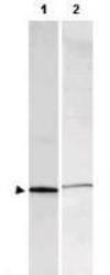
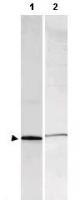
- WB using Anti-S-100 antibody shows detection of a band ~11 kDa corresponding to bovine S-100 monomer (100 ng loaded, arrowhead lane 1). The antibody also detects S-100 from rat brain lysate (lane 2). The primary antibody was diluted to 1:1,000 for 2h at RT, followed by washes and reaction with a 1:10,000 dilution of IRDye?800 conjugated Gt-a-Rabbit IgG [H&L] for 45 min at room temperature.
- Validation comment
- WB
Supportive validation
- Submitted by
- OriGene (provider)
- Main image
- Experimental details
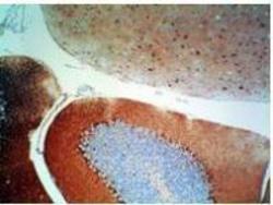
- Rabbit anti-S-100 protein was used at 1:500 to detect S-100 by IHC using a 2-step indirect method. Dark nuclear staining is observed within basket cells located near the Purkinje cells in the cerebellum. Primary antibody was diluted as stated and reacted for 30' followed by washes and the addition of donkey anti-rabbit HRP diluted 1:500 for 30'. DAB+ (Dakocytomation) was used as a substrate and was allowed to react for 5'.
- Validation comment
- IHC
- Submitted by
- OriGene (provider)
- Main image
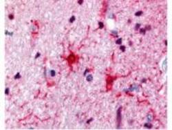
- Experimental details
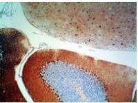
- Rabbit anti-S100 was used at a 1:500 dilution to detect S100 by immunohistochemistry in human brain astrocyte tumor tissue. Tissue was formalin-fixed and paraffin embedded. Personal Communication, Alan Yen, LifeSpanBiosciences, Seattle, WA.
- Validation comment
- IHC
