Explore
 Validate
 Learn
Learn Western blot
Antibody data
- Antibody Data
- Antigen structure
- References [0]
- Comments [0]
- Validations
- Western blot [4]
- Immunocytochemistry [2]
- Immunohistochemistry [2]
Submit
Validation data
Reference
Comment
Report error
- Product number
- PA5-27988 - Provider product page

- Provider
- Invitrogen Antibodies
- Product name
- Cyclophilin 40 Polyclonal Antibody
- Antibody type
- Polyclonal
- Antigen
- Recombinant protein fragment
- Description
- Recommended positive controls: HepG2, NIH-3T3.
- Concentration
- 0.38 mg/mL
No comments: Submit comment
Supportive validation
- Submitted by
- Invitrogen Antibodies (provider)
- Main image

- Experimental details
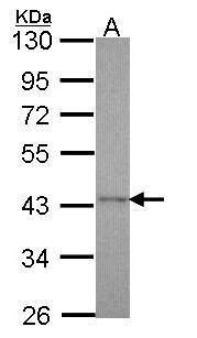
- Western Blot using Cyclophilin 40 Polyclonal Antibody (Product # PA5-27988). Sample (30 µg of whole cell lysate). A: Hep G2 . 10% SDS PAGE. Cyclophilin 40 Polyclonal Antibody (Product # PA5-27988) diluted at 1:1,000.
- Submitted by
- Invitrogen Antibodies (provider)
- Main image
- Experimental details
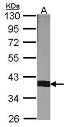
- Western Blot using Cyclophilin 40 Polyclonal Antibody (Product # PA5-27988). Sample (30 µg of whole cell lysate). A: NIH-3T3. 10% SDS PAGE. Cyclophilin 40 Polyclonal Antibody (Product # PA5-27988) diluted at 1:1,000.

- Submitted by
- Invitrogen Antibodies (provider)
- Main image

- Experimental details
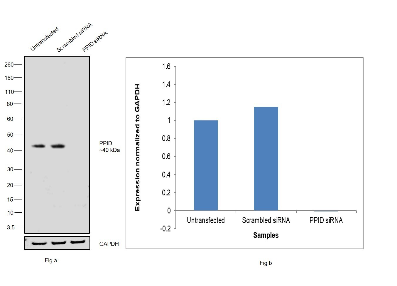
- Knockdown of Peptidylprolyl isomerase D was achieved by transfecting LNCaP with Peptidylprolyl isomerase D specific siRNAs (Silencer® select Product # S10913, S10915). Western blot analysis (Fig. a) was performed using whole cell extracts from the Peptidylprolyl isomerase D knockdown cells (lane 3), non-targeting scrambled siRNA transfected cells (lane 2) and untransfected cells (lane 1). The blot was probed with Cyclophilin 40 Polyclonal Antibody (Product # PA5-27988, 1:2000 dilution) and Goat anti-Rabbit IgG (H+L) Superclonal™ Recombinant Secondary Antibody, HRP (Product # A27036, 1:20,000 dilution). Densitometric analysis of this western blot is shown in histogram (Fig. b). Decrease in signal upon siRNA mediated knock down confirms that antibody is specific to Peptidylprolyl isomerase D.
- Submitted by
- Invitrogen Antibodies (provider)
- Main image
- Experimental details
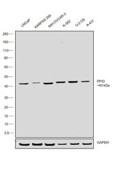
- Western blot was performed using Cyclophilin 40 Polyclonal Antibody (Product # PA5-27988) and a ~40 kDa band corresponding to Peptidylprolyl isomerase D was observed across cell lines tested. Whole cell extracts (30 µg lysate) of LNCaP (Lane 1), KARPAS 299 (Lane 2), NIH:OVCAR-3 (Lane 3), K-562 (Lane 4), U-2 OS (Lane 5) and A-431 (Lane 6) were electrophoresed using NuPAGE™ 4-12% Bis-Tris Protein Gel (Product # NP0322BOX), 12 well. Resolved proteins were then transferred onto a nitrocellulose membrane (Product # IB23001) by iBlot® 2 Dry Blotting System (Product # IB21001). The blot was probed with the primary antibody (1:2000 dilution) and detected by chemiluminescence with Goat anti-Rabbit IgG (H+L) Superclonal™ Recombinant Secondary Antibody, HRP (Product # A27036, 1:20,000 dilution) using the iBright™ FL1500 Imaging System (Product # A44115). Chemiluminescent detection was performed using SuperSignal™ West Dura Extended Duration Substrate (Product # 34076).

Supportive validation
- Submitted by
- Invitrogen Antibodies (provider)
- Main image
- Experimental details
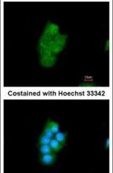
- Immunofluorescent analysis of Cyclophilin 40 in methanol-fixed HepG2 cells using a Cyclophilin 40 polyclonal antibody (Product # PA5-27988) at a 1:200 dilution.

- Submitted by
- Invitrogen Antibodies (provider)
- Main image
- Experimental details
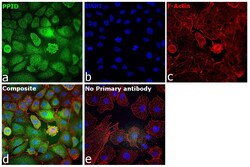
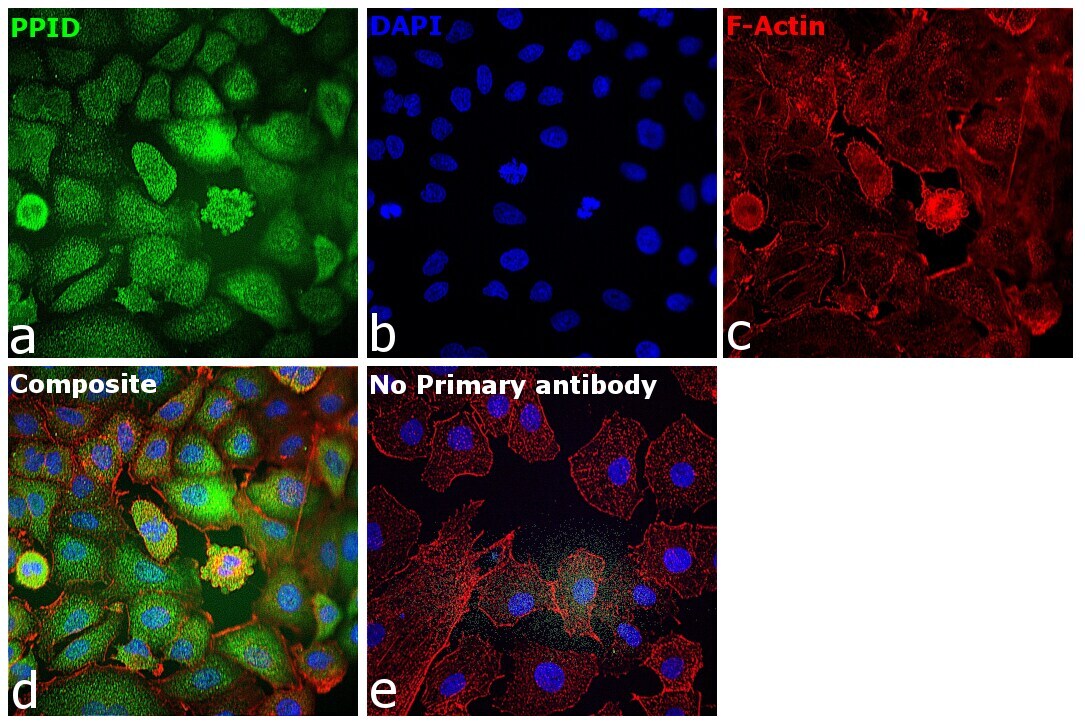
- Immunofluorescence analysis of 40 kDa Cyclophilin-40 (Peptidylprolyl isomerase D) was performed using 70% confluent log phase A549 cells. The cells were fixed with 4% paraformaldehyde for 10 minutes, permeabilized with 0.1% Triton™ X-100 for 10 minutes, and blocked with 2% BSA for overnight at room temperature. The cells were labeled with Cyclophilin 40 Polyclonal Antibody (Product # PA5-27988) at 1:100 dilution in 0.1% BSA, incubated at 4 degree celsius overnight and then labeled with Goat anti-Rabbit IgG (H+L) Superclonal™ Recombinant Secondary Antibody, Alexa Fluor® 488 conjugate (Product # A27034, 1:2000 dilution), for 45 minutes at room temperature (Panel a: Green). Nuclei (Panel b:Blue) were stained with Hoechst 33342 (Product # H1399). F-actin (Panel c: Red) was stained with Alexa Fluor™ 647 Phalloidin (Product #A22287, 1:300). Panel d represents the merged image showing Nucleus and cytoplasm localization. Panel e represents control cells with no primary antibody to assess background. The images were captured at 40x magnification in CellInsight CX7 LZR High-Content Screening (HCS) Platform (Product # CX7A1110LZR) and externally deconvoluted (D.Sage et al./Methods 115 (2017) 28–41..
Supportive validation
- Submitted by
- Invitrogen Antibodies (provider)
- Main image
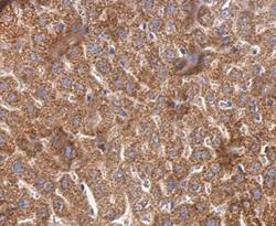
- Experimental details
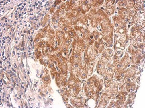
- Cyclophilin 40 Polyclonal Antibody detects Cyclophilin 40 protein at cytosol on mouse liver by immunohistochemical analysis. Sample: Paraffin-embedded mouse liver. Cyclophilin 40 Polyclonal Antibody (Product # PA5-27988) dilution: 1:500. Antigen Retrieval: EDTA based buffer, pH 8.0, 15 min.
- Submitted by
- Invitrogen Antibodies (provider)
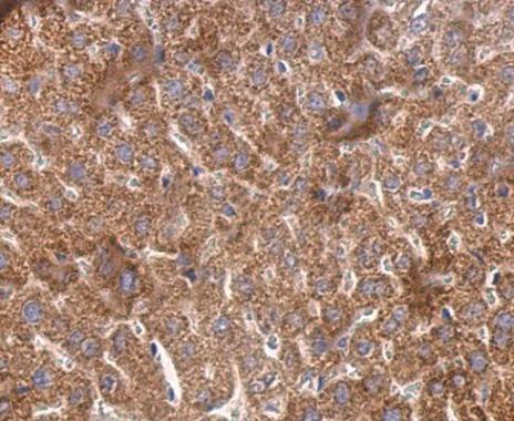
- Main image

- Experimental details
- Cyclophilin 40 Polyclonal Antibody detects PPID protein at cytosol on human hepatoma by immunohistochemical analysis. Sample: Paraffin-embedded hepatoma. Cyclophilin 40 Polyclonal Antibody (Product # PA5-27988) dilution: 1:500. Antigen Retrieval: EDTA based buffer, pH 8.0, 15 min.