Explore
 Validate
 Learn
Learn Western blot
Antibody data
- Antibody Data
- Antigen structure
- References [0]
- Comments [0]
- Validations
- Western blot [1]
- Immunocytochemistry [1]
- Immunohistochemistry [1]
Submit
Validation data
Reference
Comment
Report error
- Product number
- PA5-77516 - Provider product page

- Provider
- Invitrogen Antibodies
- Product name
- NPY1R Polyclonal Antibody
- Antibody type
- Polyclonal
- Antigen
- Synthetic peptide
- Description
- For reconstitution, we recommend adding 100 µL distilled water to a final antibody concentration of about 1 mg/mL. To use this carrier-free antibody for conjugation experiments, we strongly recommend performing another round of desalting. (Zeba Spin Desalting Columns, 7KMWCO, 0.5 mL, Product # 89882)
- Reactivity
- Human, Rat
- Host
- Rabbit
- Isotype
- IgG
- Vial size
- 50 µL
- Concentration
- 0.75 mg/mL
- Storage
- -20°C
No comments: Submit comment
Supportive validation
- Submitted by
- Invitrogen Antibodies (provider)
- Main image

- Experimental details
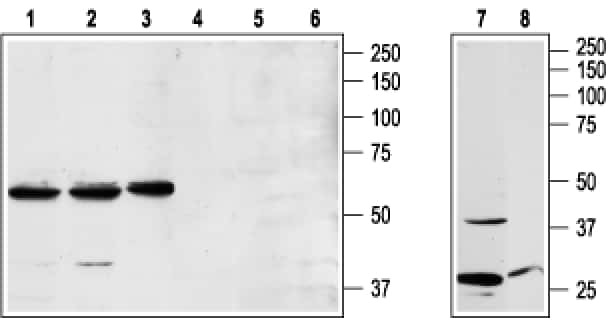
- Western blot analysis of Jurkat (lanes 1 and 4), K562 (lanes 2 and 5), and RBL (lanes 3 and 6) cell lysates and rat brain lysates (lanes 7 and 8) with NPY1R polyclonal antibody (Product # PA5-77516) using a dilution of 1:200.
Supportive validation
- Submitted by
- Invitrogen Antibodies (provider)
- Main image
- Experimental details
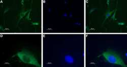
- Immunocytochemistry analysis of NPY1R in paraformaldehyde-fixed and permeabilized rat dorsal root ganglion. A, D) Samples were probed with NPY1R polyclonal antibody (Product # PA5-77516) at a dilution of 1:100, and incubated with goat-anti-rabbit-AlexaFluor-555 and Hoechst. B, E) Shows nuclei stained image. C) Shows merged images of Panels A and B. F) Shows merged images of Panels D and E. Magnification: A-C: x20, E-F:x100.

Supportive validation
- Submitted by
- Invitrogen Antibodies (provider)
- Main image

- Experimental details
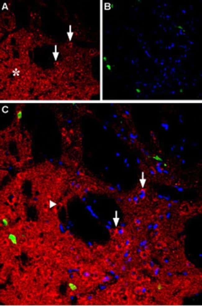
- Immunohistochemistry analysis of NPY1R in rat striatum. A) Samples were probed with NPY1R polyclonal antibody (Product # PA5-77516) and incubated with parvalbumin (green) and DAPI. B) Image showing striatal matrix. C) Merged image.