Explore
 Validate
 Learn
Learn Western blot
Antibody data
- Antibody Data
- Antigen structure
- References [0]
- Comments [0]
- Validations
- Western blot [1]
- Flow cytometry [1]
Submit
Validation data
Reference
Comment
Report error
- Product number
- MA5-24108 - Provider product page

- Provider
- Invitrogen Antibodies
- Product name
- NAMPT Monoclonal Antibody (362616)
- Antibody type
- Monoclonal
- Antigen
- Recombinant full-length protein
- Description
- In direct ELISAs and Western blots, 100% cross-reactivity with recombinant human PBEF is observed.
- Antibody clone number
- 362616
- Concentration
- 0.5 mg/mL
No comments: Submit comment
Supportive validation
- Submitted by
- Invitrogen Antibodies (provider)
- Main image
- Experimental details
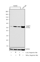
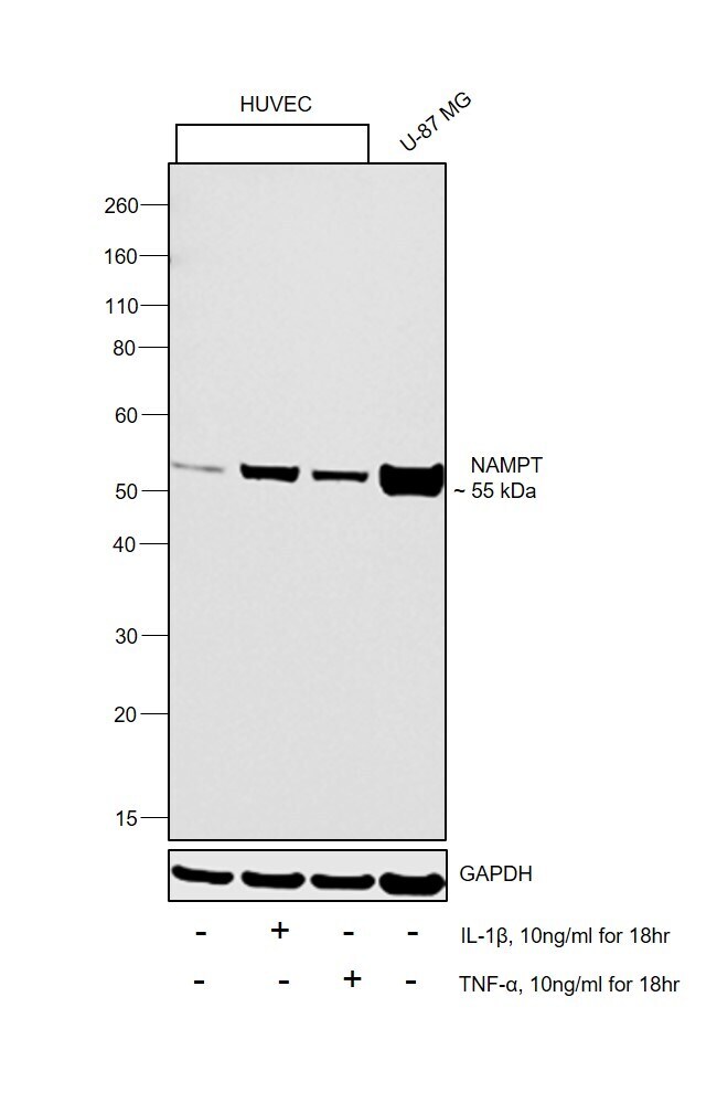
- Western blot was performed using Anti-NAMPT Monoclonal Antibody (362616)(Product # MA5-24108) and a 55 kDa band corresponding to NAMPT was observed across cell lines tested. Increased expression of NAMPT was observed in HUVEC upon IL1-beta and TNF-alpha treatment. Whole cell extracts (30 µg lysate) of HUVEC (Lane 1), HUVEC treated with IL1-beta (10 ng/mL for 18hr) (Lane 2), HUVEC treated with TNF-alpha (10 ng/mL for 18hr) (Lane 3) and U-87 MG (Lane 4) were electrophoresed using NuPAGE™ 4-12% Bis-Tris Protein Gel (Product # NP0322BOX). Resolved proteins were then transferred onto a Nitrocellulose membrane (Product # IB23001) by iBlot® 2 Dry Blotting System (Product # IB21001). The blot was probed with the primary antibody (1 µg/mL) and detected by chemiluminescence with F(ab2-Rabbit anti-Rat IgG (H+L Secondary Antibody, HRP (Product # PA1-29927,1:4000 dilution) using the iBright FL 1000 (Product # A32752). Chemiluminescent detection was performed using Novex® ECL Chemiluminescent Substrate Reagent Kit (Product # WP20005).
Supportive validation
- Submitted by
- Invitrogen Antibodies (provider)
- Main image

- Experimental details
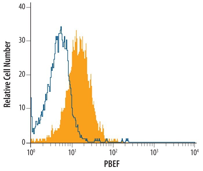
- Flow cytometry of NAMPT in 3T3‚L1 mouse embryonic fibroblast adipose‚like cell line. Samples were incubated in NAMPT monoclonal antibody (Product # MA5-24108) or isotype control antibody followed by Allophycocyanin-conjugated Anti-Rat IgG F(ab)2 Secondary Antibody. To facilitate intracellular staining, cells were fixed with paraformaldehyde and permeabilized with saponin.