 Explore
 Validate
 Learn
Learn Western blot
Antibody data
- Antibody Data
- Antigen structure
- References [0]
- Comments [0]
- Validations
- Western blot [3]
- Immunohistochemistry [1]
Submit
Validation data
Reference
Comment
Report error
- Product number
- PA5-64984 - Provider product page

- Provider
- Invitrogen Antibodies
- Product name
- PTGES2 Polyclonal Antibody
- Antibody type
- Polyclonal
- Antigen
- Recombinant full-length protein
- Description
- Immunogen sequence: AQDLHAERSAA QLSLSSRLQL TLYQYKTCPF CSKVRAFLDF HALPYQVVEV NPVRRAEIKF SSYRKVPILV AQEGESSQ
- Concentration
- 0.05 mg/mL
No comments: Submit comment
Supportive validation
- Submitted by
- Invitrogen Antibodies (provider)
- Main image

- Experimental details
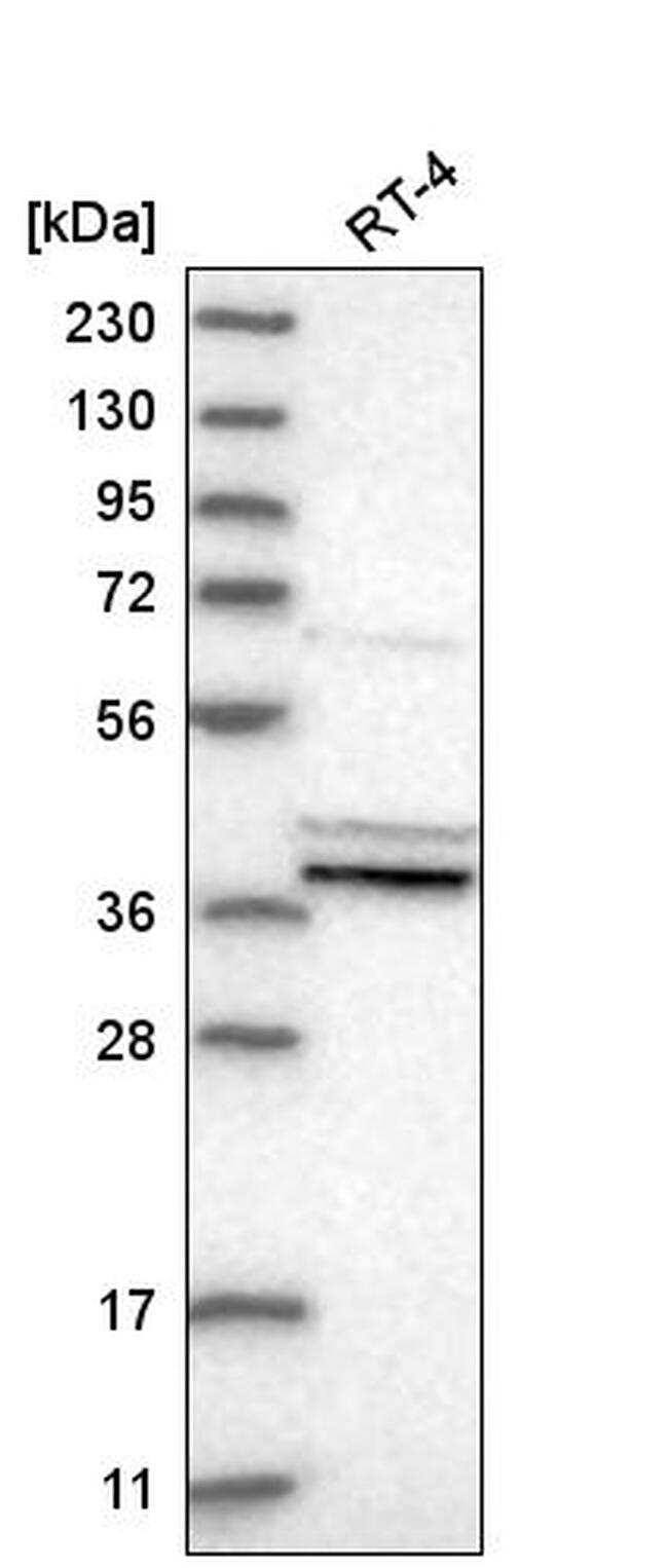
- Western blot analysis of PTGES2 in human cell line RT-4. Samples were probed using a PTGES2 Polyclonal Antibody (Product # PA5-64984).
- Submitted by
- Invitrogen Antibodies (provider)
- Main image

- Experimental details
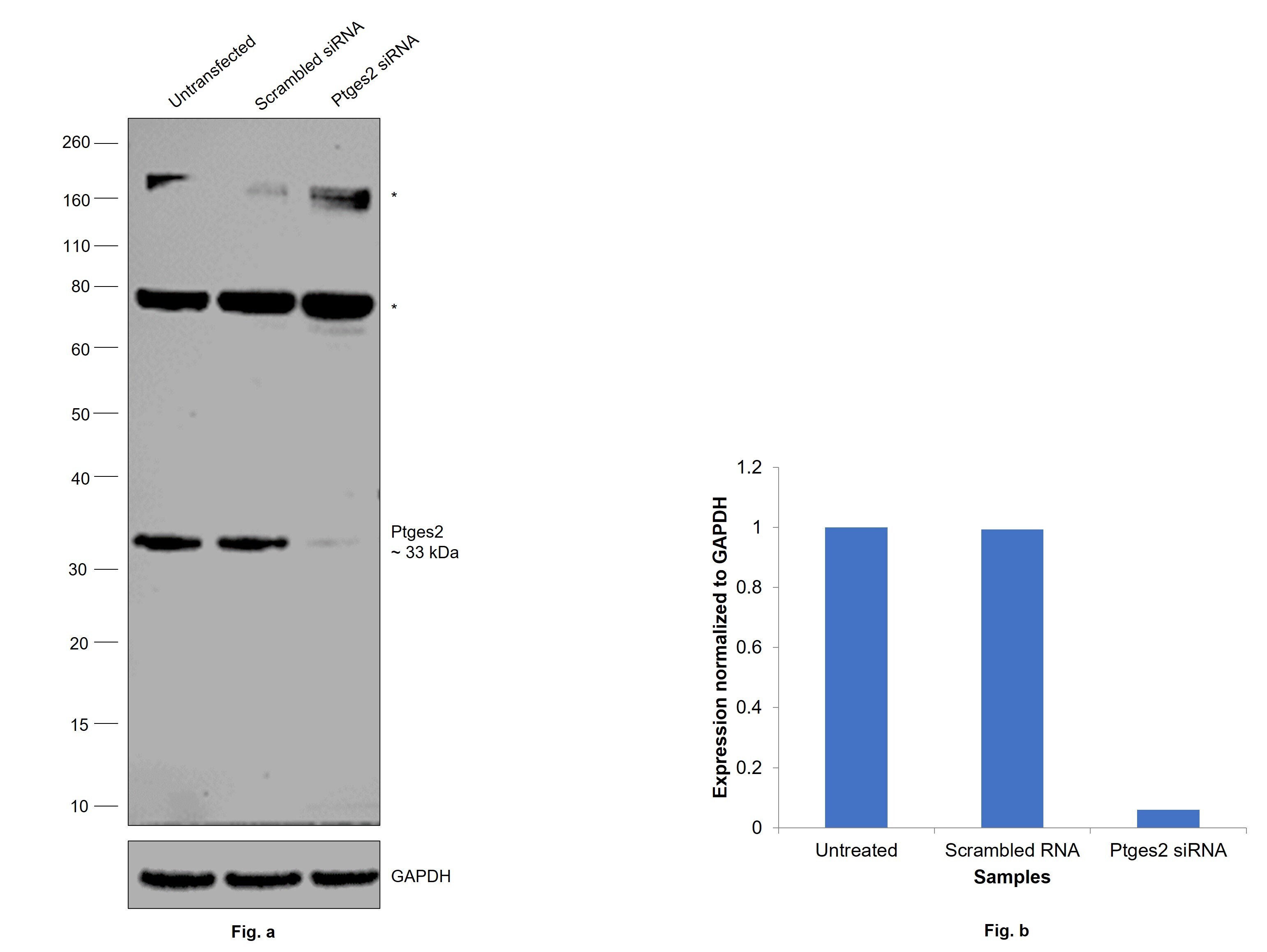
- Knockdown of Prostaglandin E synthase 2 was achieved by transfecting SW480 with Prostaglandin E synthase 2 specific siRNAs (Silencer® select Product # S36951, S36953). Western blot analysis (Fig. a) was performed using Membrane enriched extracts from Prostaglandin E synthase 2 knockdown cells (lane 3), non-targeting scrambled siRNA transfected cells (lane 2) and untransfected cells (lane 1). The blot was probed with PTGES2 Polyclonal Antibody (Product # PA5-64984, 0.2 µg/mL) and Goat anti-Rabbit IgG (H+L) Superclonal™ Recombinant Secondary Antibody, HRP (Product # A27036, 1:20,000 dilution). Densitometric analysis of this western blot is shown in histogram (Fig. b). Decrease in signal upon siRNA mediated knock down confirms that antibody is specific to Prostaglandin E synthase 2. Uncharacterized bands (*) were observed at ~80 kDa and ~180 kDa in all the lysates.
- Submitted by
- Invitrogen Antibodies (provider)
- Main image
- Experimental details
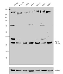
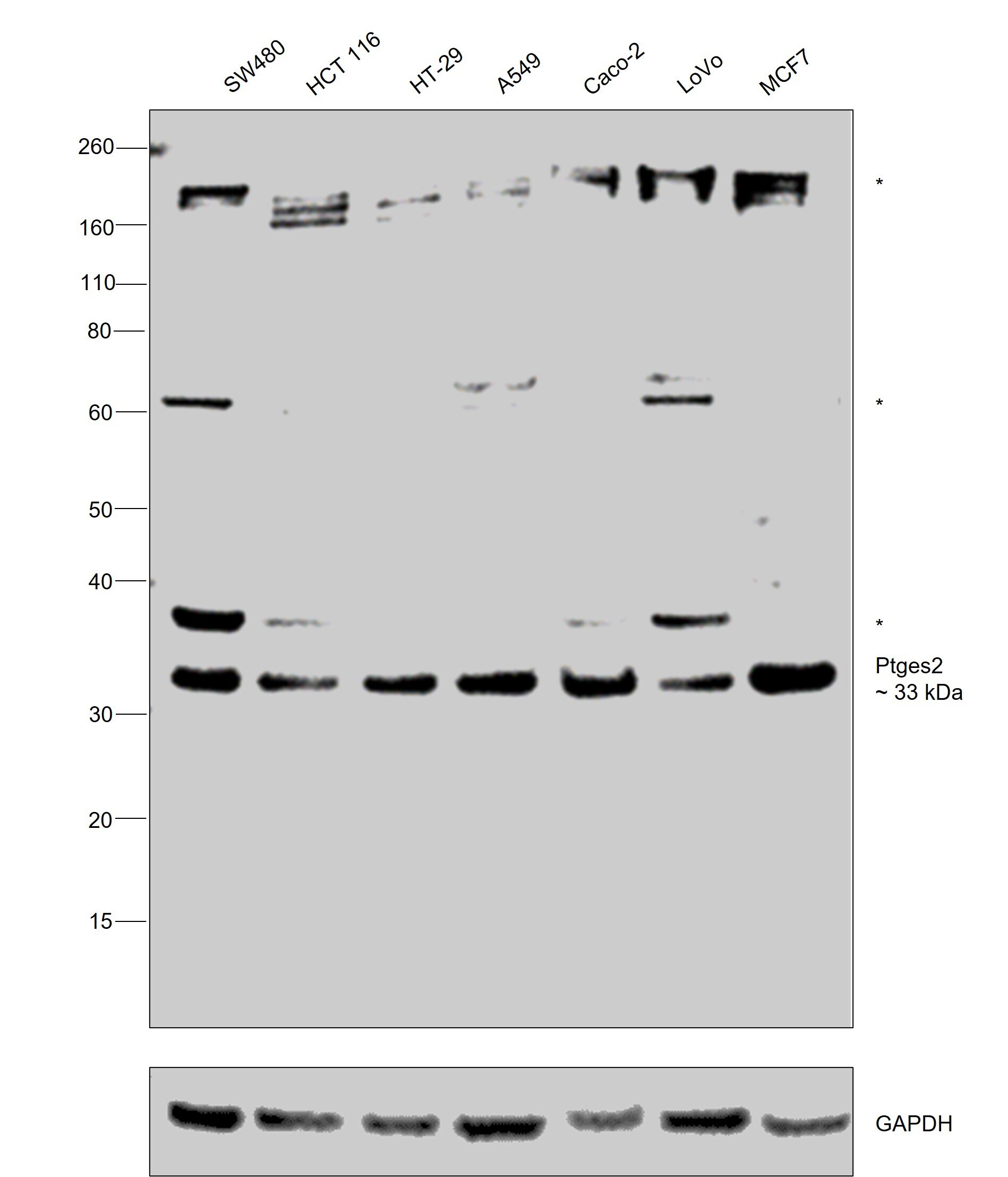
- Western blot was performed using Anti-PTGES2 Polyclonal Antibody (Product # PA5-64984) and a 33 kDa band corresponding to Prostaglandin E synthase 2 was observed across all cell lysates tested. Membrane enriched extracts (30 µg lysate) of SW480 (Lane 1), HCT 116 (Lane 2), HT-29 (Lane 3), A549 (Lane 4), Caco-2 (Lane 5), LoVo (Lane 6) and MCF7 (Lane 7) were electrophoresed using NuPAGE™ 4-12% Bis-Tris Protein Gel (Product # NP0322BOX), 12 well. Resolved proteins were then transferred onto a nitrocellulose membrane (Product # IB23001) by iBlot® 2 Dry Blotting System (Product # IB21001). The blot was probed with the primary antibody (0.2 µg/mL) and detected by chemiluminescence with Goat anti-Rabbit IgG (H+L) Superclonal™ Recombinant Secondary Antibody, HRP (Product # A27036, 1:20,000 dilution) using the iBright™ FL1500 Imaging System (Product # A44115). Chemiluminescent detection was performed using SuperSignal™ West Atto Ultimate Sensitivity Substrate (Product # A38556). Uncharacterized bands (*) were observed at ~38 kDa, ~62 kDa in some cell lysates and all cell lysates displayed an uncharacterized band (*) at ~180 kDa.
Supportive validation
- Submitted by
- Invitrogen Antibodies (provider)
- Main image
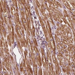
- Experimental details
- Immunohistochemical staining of PTGES2 in human heart muscle shows strong cytoplasmic positivity in myocytes. Samples were probed using a PTGES2 Polyclonal Antibody (Product # PA5-64984).
