Explore
 Validate
 Learn
Learn Western blot
 Immunoprecipitation
Antibody data
- Antibody Data
- Antigen structure
- References [0]
- Comments [0]
- Validations
- Western blot [2]
- Immunohistochemistry [2]
- Flow cytometry [1]
Submit
Validation data
Reference
Comment
Report error
- Product number
- MA5-16184 - Provider product page

- Provider
- Invitrogen Antibodies
- Product name
- TLR3 Monoclonal Antibody (40C1285.6)
- Antibody type
- Monoclonal
- Antigen
- Synthetic peptide
- Reactivity
- Human, Mouse, Canine
- Host
- Mouse
- Isotype
- IgG
- Antibody clone number
- 40C1285.6
- Vial size
- 100 µg
- Concentration
- 1 mg/mL
- Storage
- Store at 4°C short term. For long term storage, store at -20°C, avoiding freeze/thaw cycles.
No comments: Submit comment
Supportive validation
- Submitted by
- Invitrogen Antibodies (provider)
- Main image
- Experimental details
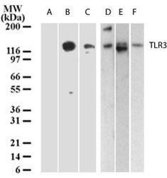
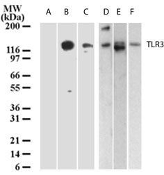
- Western blot analysis of TLR3 in lysates from A) untransfected 293, B) 293 cells transfected with human TLR3 cDNA, C) human intestine, D) placenta, E) heart and F) ovary using a TLR3/CD283 monoclonal antibody (Product # MA5-16184) at 3 µg/mL.
- Submitted by
- Invitrogen Antibodies (provider)
- Main image
- Experimental details
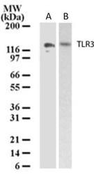
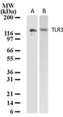
- Western blot analysis of TLR3 in lysates from human intestine and ovary. Samples were incubated in TLR3 monoclonal antibody (Product # MA5-16184) using a dilution of 3 µg/mL followed by a goat anti-mouse HRP secondary antibody.
Supportive validation
- Submitted by
- Invitrogen Antibodies (provider)
- Main image
- Experimental details
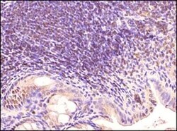
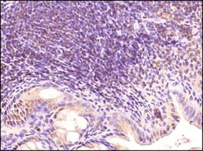
- Immunohistochemical analysis of TLR3 in Tissue section of mouse intestine. Samples were incubated in TLR3 monoclonal antibody (Product # MA5-16184) using a dilution of 1:500 followed by HRP-DAB detection and hematoxylin counterstaining. The representative image shows a punctate staining of the ER and endosomes in a subset of cells in Peyers patches (organized lymphoid nodules) in the tested section.
- Submitted by
- Invitrogen Antibodies (provider)
- Main image
- Experimental details
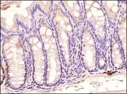
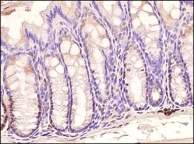
- Immunohistochemical analysis of TLR3 in Mouse colon. Samples were incubated in TLR3 monoclonal antibody (Product # MA5-16184) using a dilution of 1:500 followed by HRP-DAB detection and hematoxylin counterstaining. Intense signal was found in subset of cells at the bases of the crypts.
Supportive validation
- Submitted by
- Invitrogen Antibodies (provider)
- Main image
- Experimental details
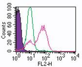
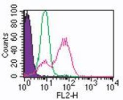
- Intracellular flow analysis of TLR3 in human monocytes using 0.5 µg of TLR3/CD283 monoclonal antibody (Product # MA5-16184). Shaded histogram represents monocytes without antibody; green represents isotype control; red represents TLR3/CD283 monoclonal antibody (Product # MA5-16184). Goat anti-mouse IgG1 PE conjugate was used as secondary.