 Explore
 Validate
 Learn
Learn Western blot
Antibody data
- Antibody Data
- Antigen structure
- References [1]
- Comments [0]
- Validations
- Western blot [4]
- Immunocytochemistry [2]
- Immunohistochemistry [4]
Submit
Validation data
Reference
Comment
Report error
- Product number
- PA5-20183 - Provider product page

- Provider
- Invitrogen Antibodies
- Product name
- TLR3 Polyclonal Antibody
- Antibody type
- Polyclonal
- Antigen
- Synthetic peptide
- Description
- A suggested positive control is Daudi cell lysate.
- Concentration
- 1 mg/mL
Submitted references Protective action of Bacillus clausii probiotic strains in an in vitro model of Rotavirus infection.
Paparo L, Tripodi L, Bruno C, Pisapia L, Damiano C, Pastore L, Berni Canani R
Scientific reports 2020 Jul 28;10(1):12636
Scientific reports 2020 Jul 28;10(1):12636
No comments: Submit comment
Supportive validation
- Submitted by
- Invitrogen Antibodies (provider)
- Main image
- Experimental details
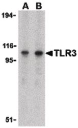
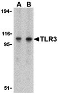
- Western blot analysis of Daudi cell lysate using a CD283/TLR3/Toll-like Receptor 3 polyclonal antibody (Product # PA5-20183) at (A) 1 and (B) 2 µg/mL.
- Submitted by
- Invitrogen Antibodies (provider)
- Main image

- Experimental details
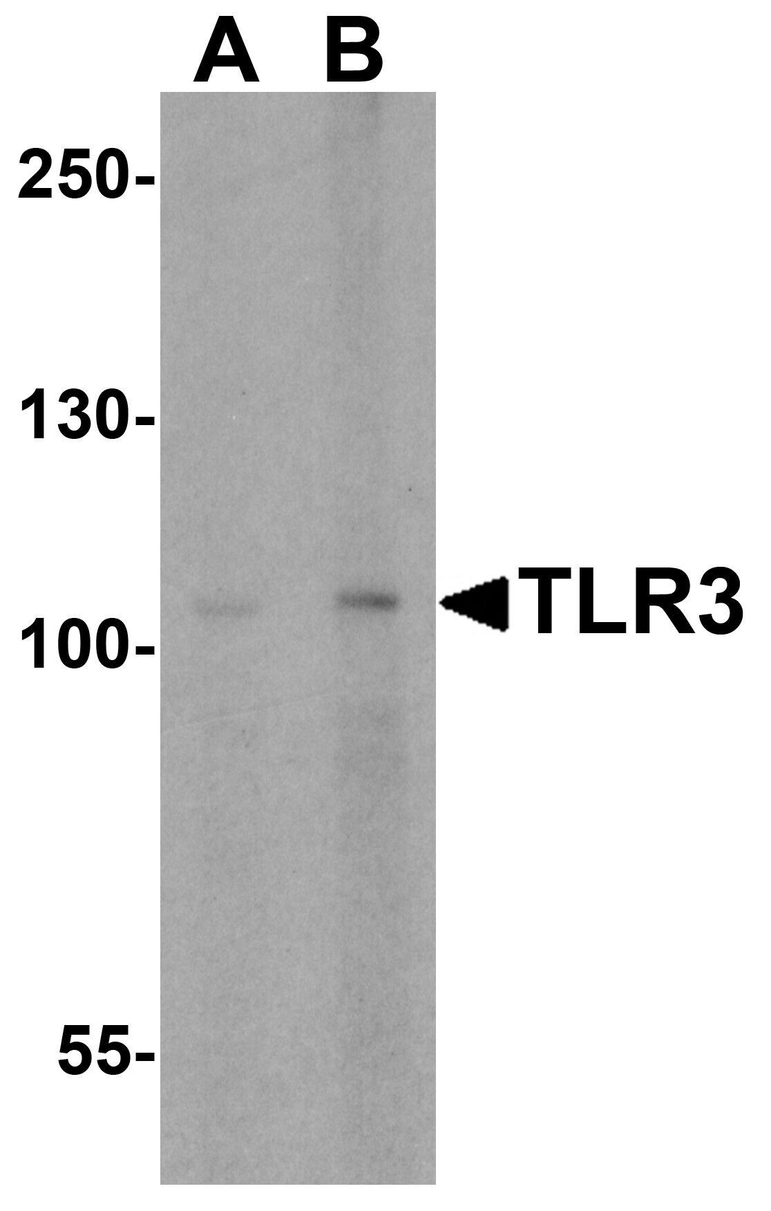
- Western Blot analysis of TLR3 expression in mouse spleen tissue lysate with TLR3 Polyclonal Antibody (Product # PA5-20183) at (A) 0.5 and (B) 1 µg/mL.
- Submitted by
- Invitrogen Antibodies (provider)
- Main image

- Experimental details
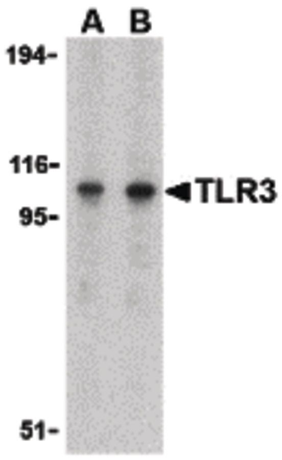
- Western Blot analysis of TLR3 in Daudi cell lysate with TLR3 Polyclonal Antibody (Product # PA5-20183) at (A) 1 and (B) 2 µg/mL.
- Submitted by
- Invitrogen Antibodies (provider)
- Main image
- Experimental details
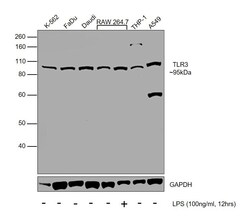
- Western blot was performed using Anti-TLR3 Polyclonal Antibody (Product # PA5-20183) and a 95kDa band corresponding to TLR3 was observed in all the tested cell models, and was upregulated upon LPS treatment in RAW 264.7. Whole cell lysate (30ug lysate) of K562 (Lane 1), FaDu (Lane 2), Daudi (Lane 3), RAW 264.7 (Lane 4), RAW 264.7 treated with LPS (100ng/ml for 12hrs) (Lane 5), THP-1 (Lane 6) and A549 (Lane 7) were electrophoresed using NuPAGE® 10 % Bis-Tris gel (Product # NP0302BOX). Resolved proteins were then transferred onto a nitrocellulose membrane (Product # IB23001) by iBlot® 2 Dry Blotting System (Product # IB21001). The blot was probed with the primary antibody (1µg/ml concentration) and detected by chemiluminescence with Goat anti-Rabbit IgG (H+L) Superclonal™ Recombinant Secondary Antibody, HRP (Product # A27036, 1:4000 dilution) using the iBright FL 1000 (Product # A32752). Chemiluminescent detection was performed using Novex® ECL Chemiluminescent Substrate Reagent Kit (Product # WP20005).
Supportive validation
- Submitted by
- Invitrogen Antibodies (provider)
- Main image
- Experimental details
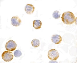
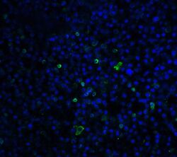
- Immunocytochemistry of TLR3 in EL4 cells with TLR3 Polyclonal Antibody (Product # PA5-20183) at 1 µg/mL.

- Submitted by
- Invitrogen Antibodies (provider)
- Main image
- Experimental details
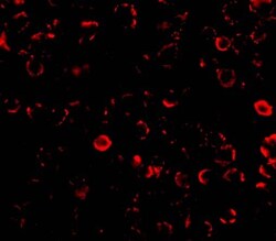
- Immunofluorescence of TLR3 in El4 cells with TLR3 Polyclonal Antibody (Product # PA5-20183) at 10 µg/mL.
Supportive validation
- Submitted by
- Invitrogen Antibodies (provider)
- Main image
- Experimental details
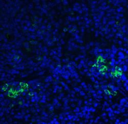
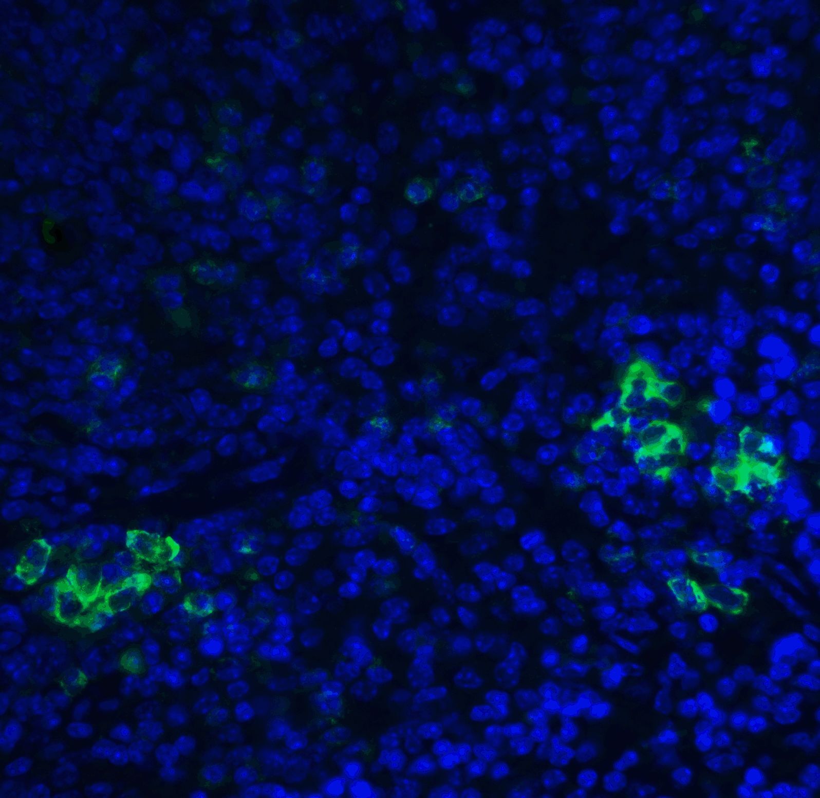
- Immunofluorescence of TLR3 in human spleen tissue with TLR3 Polyclonal Antibody (Product # PA5-20183) at 20 µg/mL. Green: TLR3 Blue: DAPI staining
- Submitted by
- Invitrogen Antibodies (provider)
- Main image
- Experimental details
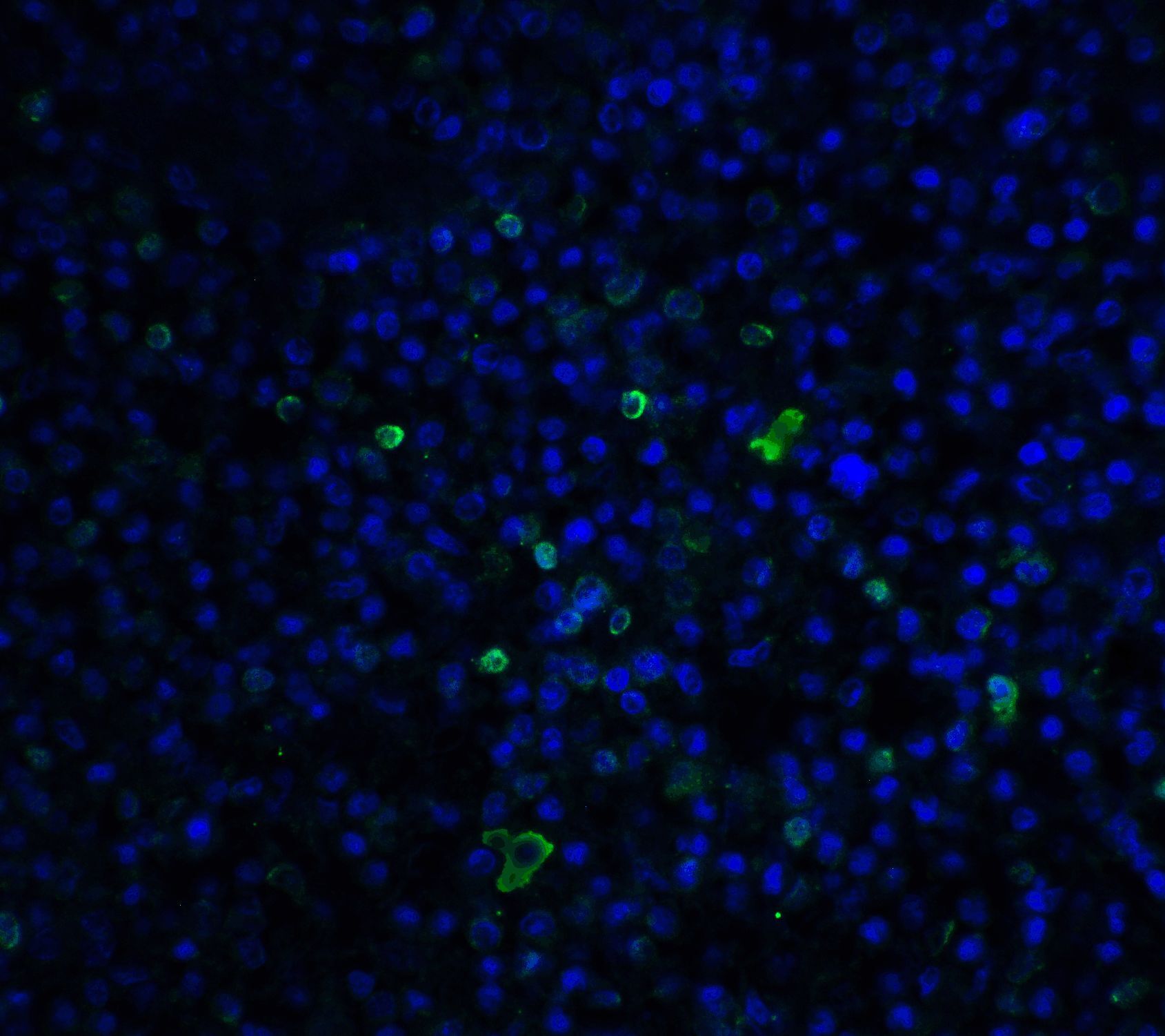
- Immunofluorescence of TLR3 in mouse spleen tissue with TLR3 Polyclonal Antibody (Product # PA5-20183) at 20 µg/mL. Green: TLR3 Blue: DAPI staining
- Submitted by
- Invitrogen Antibodies (provider)
- Main image
- Experimental details
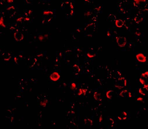
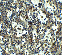
- Immunohistochemistry of TLR3 in human spleen tissue with TLR3 Polyclonal Antibody (Product # PA5-20183) at 5 µg/mL.

- Submitted by
- Invitrogen Antibodies (provider)
- Main image
- Experimental details
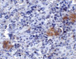
- Immunohistochemistry of TLR3 in mouse spleen tissue with TLR3 Polyclonal Antibody (Product # PA5-20183) at 2 µg/mL.
