Explore
 Validate
 Learn
Learn Western blot
Antibody data
- Antibody Data
- Antigen structure
- References [0]
- Comments [0]
- Validations
- Western blot [4]
- Immunocytochemistry [1]
- Immunohistochemistry [9]
Submit
Validation data
Reference
Comment
Report error
- Product number
- MA5-27369 - Provider product page

- Provider
- Invitrogen Antibodies
- Product name
- hnRNP H1 Monoclonal Antibody (OTI2E8)
- Antibody type
- Monoclonal
- Antigen
- Recombinant full-length protein
- Reactivity
- Human
- Host
- Mouse
- Isotype
- IgG
- Antibody clone number
- OTI2E8
- Vial size
- 100 µL
- Concentration
- 1 mg/mL
- Storage
- -20° C, Avoid Freeze/Thaw Cycles
No comments: Submit comment
Supportive validation
- Submitted by
- Invitrogen Antibodies (provider)
- Main image
- Experimental details
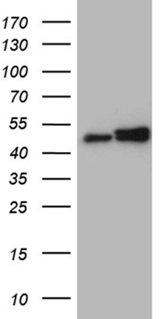
- Western blot analysis of HNRNPH1 in HEK293T cells in untransfected (Left lane) and transfected (Right lane) samples using 5 µg per lane. The samples were separated by SDS-PAGE and probed with HNRNPH1 (Product # MA5-27369) monoclonal antibody.
- Submitted by
- Invitrogen Antibodies (provider)
- Main image
- Experimental details
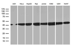
- Western blot analysis of HNRNPH1 in A431, HeLa, HepG2, RAJI, Jurkat, K562, U251, HUH7 cells using 35 µg per lane. Samples were probed with HNRNPH1 (Product # MA5-27369) monoclonal antibody at a dilution of 1:500.

- Submitted by
- Invitrogen Antibodies (provider)
- Main image
- Experimental details
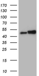
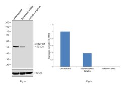
- Knockdown of hnRNP H1 was achieved by transfecting Hep G2 with hnRNP H1 specific siRNAs (Silencer® select Product # s6728, s6729). Western blot analysis (Fig. a) was performed using modified whole cell extracts (1%SDS) from the knockdown cells (Lane 3), non-specific scrambled siRNA transfected cells (Lane 2) and untransfected cells (Lane 1). The blot was probed with hnRNP H1 Monoclonal Antibody (OTI2E8) (Product # MA5-27369, 1:1000 dilution) and Goat anti-Mouse IgG (H+L), Superclonal™ Recombinant Secondary Antibody, HRP conjugate (Product # A28177, 0.25µg/mL, 1:4000 dilution). Densitometric analysis of this western blot is shown in histogram (Fig. b). Decrease in signal upon siRNA mediated knock down confirms that antibody is specific to hnRNP H1.

- Submitted by
- Invitrogen Antibodies (provider)
- Main image

- Experimental details
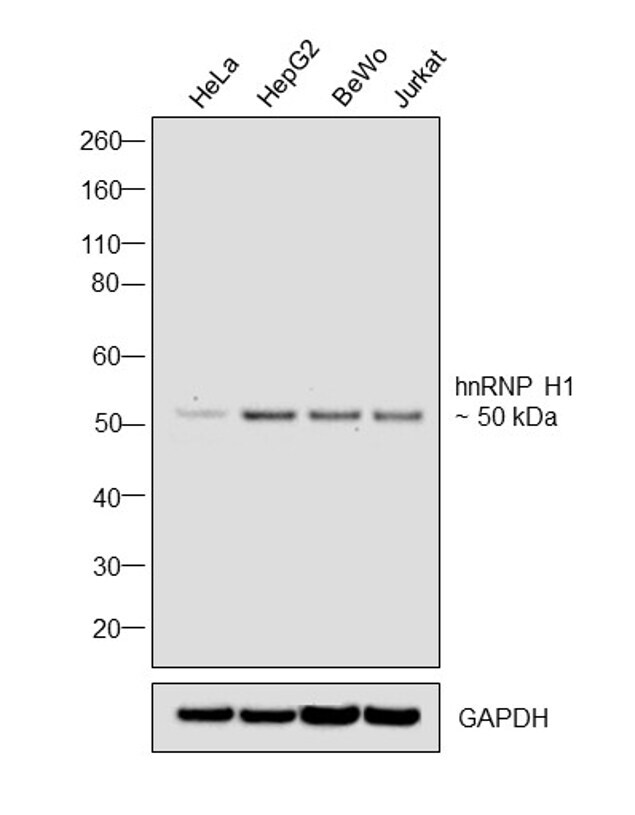
- Western blot was performed using Anti-hnRNP H1 Monoclonal Antibody (OTI2E8), (Product # MA5-27369) and a 50 kDa band corresponding to hnRNP H1 was observed in the cell lines tested. Modified whole cell extracts (1%SDS) (30 µg lysate) of HeLa (Lane 1), Hep G2 (Lane 2), BeWo (Lane 3), and Jurkat (Lane 4) were electrophoresed using Novex® NuPAGE® 4-12 % Bis-Tris gel (Product # NP0321BOX). Resolved proteins were then transferred onto a nitrocellulose membrane (Product # IB23001) by iBlot® 2 Dry Blotting System (Product # IB21001). The blot was probed with the primary antibody (1:1000 dilution) and detected by chemiluminescence with Goat anti-Mouse IgG (H+L), Superclonal™ Recombinant Secondary Antibody, HRP (Product # A28177, 1:4000 dilution) using the iBright FL 1000 (Product # A32752). Chemiluminescent detection was performed using Novex® ECL Chemiluminescent Substrate Reagent Kit (Product # WP20005).
Supportive validation
- Submitted by
- Invitrogen Antibodies (provider)
- Main image
- Experimental details
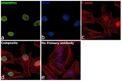
- Immunofluorescence analysis of HNRNPH1 was performed using 70% confluent log phase HeLa cells. The cells were fixed with 4% paraformaldehyde for 10 minutes, permeabilized with 0.1% Triton™ X-100 for 15 minutes, and blocked with 2% BSA for 1 hour at room temperature. The cells were labeled with hnRNP H1 Monoclonal Antibody (OTI2E8) (Product # MA5-27369) at 1:200 dilution in 0.1% BSA, incubated at 4 degree celsius overnight and then with Donkey anti-Mouse IgG (H+L) Highly Cross-Adsorbed Secondary Antibody, Alexa Fluor Plus 488 (Product # A32766) at a dilution of 1:2000 for 45 minutes at room temperature (Panel a: green). Nuclei (Panel b: blue) were stained with SlowFade® Gold Antifade Mountant with DAPI (Product # S36938). F-actin (Panel c: red) was stained with Rhodamine Phalloidin (Product # R415, 1:300). Panel d represents the merged image showing staining in nucleus. Panel e represents control cells with no primary antibody to assess background. The images were captured at 60X magnification.

Supportive validation
- Submitted by
- Invitrogen Antibodies (provider)
- Main image
- Experimental details
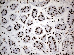
- Immunohistochemistry was performed on paraffin-embedded human gastric tissue. To expose target proteins, heat-induced epitope retrieval by 10mM citric buffer, pH6.0, 100°C for 10min. Following antigen retrieval, tissues were probed with a HNRNPH1 monoclonal antibody (Product # MA5-27369) at a dilution of 1:500.

- Submitted by
- Invitrogen Antibodies (provider)
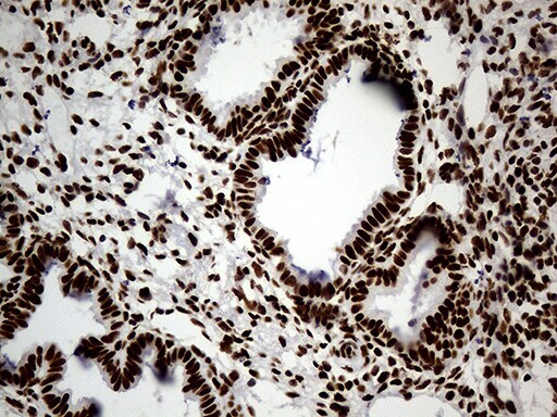
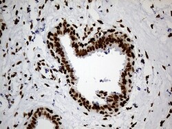
- Main image
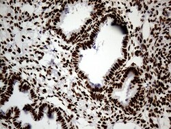
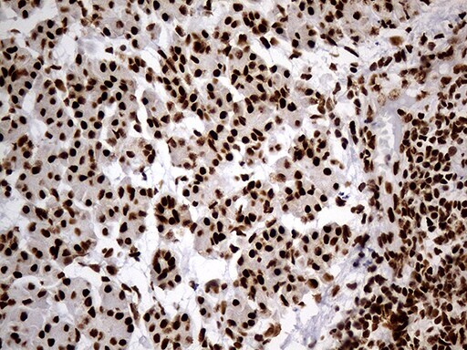
- Experimental details
- Immunohistochemistry was performed on paraffin-embedded human endometrium tissue. To expose target proteins, heat-induced epitope retrieval by Tris-EDTA. Following antigen retrieval, tissues were probed with a HNRNPH1 monoclonal antibody (Product # MA5-27369) at a dilution of 1:500.
- Submitted by
- Invitrogen Antibodies (provider)
- Main image
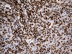
- Experimental details
- Immunohistochemistry was performed on paraffin-embedded human spleen tissue. To expose target proteins, heat-induced epitope retrieval by 10mM citric buffer, pH6.0, 100°C for 10min. Following antigen retrieval, tissues were probed with a HNRNPH1 monoclonal antibody (Product # MA5-27369) at a dilution of 1:500.

- Submitted by
- Invitrogen Antibodies (provider)
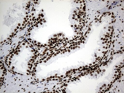
- Main image
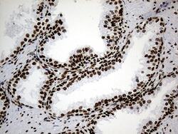
- Experimental details
- Immunohistochemistry was performed on paraffin-embedded human prostate tissue. To expose target proteins, heat-induced epitope retrieval by Tris-EDTA. Following antigen retrieval, tissues were probed with a HNRNPH1 monoclonal antibody (Product # MA5-27369) at a dilution of 1:500.
- Submitted by
- Invitrogen Antibodies (provider)
- Main image
- Experimental details
- Immunohistochemistry was performed on paraffin-embedded human breast tissue. To expose target proteins, heat-induced epitope retrieval by 10mM citric buffer, pH6.0, 100°C for 10min. Following antigen retrieval, tissues were probed with a HNRNPH1 monoclonal antibody (Product # MA5-27369) at a dilution of 1:500.

- Submitted by
- Invitrogen Antibodies (provider)
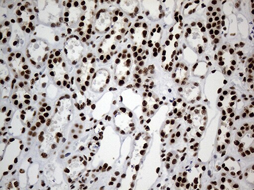
- Main image
- Experimental details
- Immunohistochemistry was performed on paraffin-embedded human kidney tissue. To expose target proteins, heat-induced epitope retrieval by 10mM citric buffer, pH6.0, 100°C for 10min. Following antigen retrieval, tissues were probed with a HNRNPH1 monoclonal antibody (Product # MA5-27369) at a dilution of 1:500.
- Submitted by
- Invitrogen Antibodies (provider)
- Main image
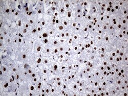
- Experimental details
- Immunohistochemistry was performed on paraffin-embedded human liver tissue. To expose target proteins, heat-induced epitope retrieval by 10mM citric buffer, pH6.0, 100°C for 10min. Following antigen retrieval, tissues were probed with a HNRNPH1 monoclonal antibody (Product # MA5-27369) at a dilution of 1:500.

- Submitted by
- Invitrogen Antibodies (provider)
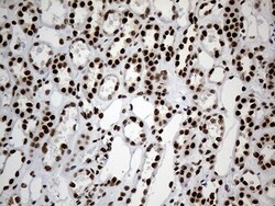
- Main image

- Experimental details
- Immunohistochemistry was performed on paraffin-embedded human pancreas tissue. To expose target proteins, heat-induced epitope retrieval by 10mM citric buffer, pH6.0, 100°C for 10min. Following antigen retrieval, tissues were probed with a HNRNPH1 monoclonal antibody (Product # MA5-27369) at a dilution of 1:500.
- Submitted by
- Invitrogen Antibodies (provider)
- Main image
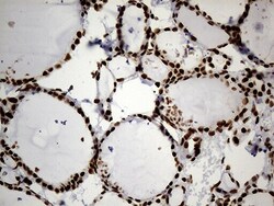
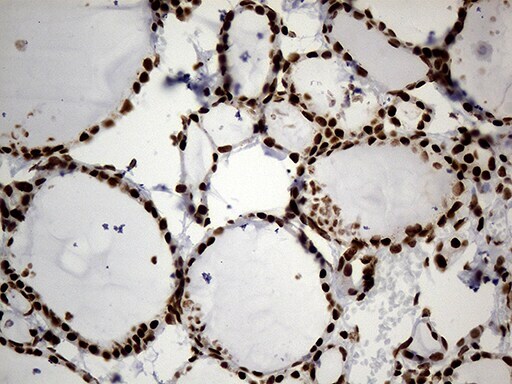
- Experimental details
- Immunohistochemistry was performed on paraffin-embedded human thyroid tissue. To expose target proteins, heat-induced epitope retrieval by 10mM citric buffer, pH6.0, 100°C for 10min. Following antigen retrieval, tissues were probed with a HNRNPH1 monoclonal antibody (Product # MA5-27369) at a dilution of 1:500.