Explore
 Validate
 Learn
Learn Western blot
 Immunocytochemistry
Antibody data
- Antibody Data
- Antigen structure
- References [0]
- Comments [0]
- Validations
- Western blot [6]
- Immunocytochemistry [1]
Submit
Validation data
Reference
Comment
Report error
- Product number
- GTX630397 - Provider product page

- Provider
- GeneTex
- Product name
- NDP52 antibody [GT1813]
- Antibody type
- Monoclonal
- Reactivity
- Human
- Host
- Mouse
No comments: Submit comment
Enhanced validation
Supportive validation
- Submitted by
- GeneTex (provider)
- Enhanced method
- Genetic validation
- Main image
- Experimental details
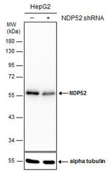
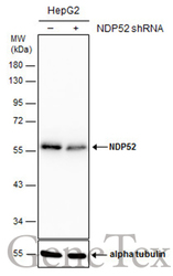
- Non-transfected (¡V) and transfected (+) HepG2 whole cell extracts (30 ?g) were separated by 10% SDS-PAGE, and the membrane was blotted with NDP52 antibody [GT1813] (GTX630397) diluted at 1:1000.
Supportive validation
- Submitted by
- GeneTex (provider)
- Main image

- Experimental details
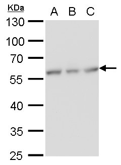
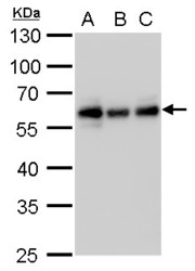
- NDP52 antibody [GT1813] detects NDP52 protein by western blot analysis.A. 30 £gg Huh7 whole cell lysate/extract B. 30 £gg Hep3B whole cell lysate/extract C. 30 £gg HepG2 whole cell lysate/extract10 % SDS-PAGENDP52 antibody [GT1813] (GTX630397) dilution: 1:1000
- Submitted by
- GeneTex (provider)
- Main image
- Experimental details
- NDP52 antibody [GT1813] detects NDP52 protein by western blot analysis.A. 30 £gg Jurkat whole cell lysate/extract B. 30 £gg Raji whole cell lysate/extract C. 30 £gg NCI-H929 whole cell lysate/extract10 % SDS-PAGENDP52 antibody [GT1813] (GTX630397) dilution: 1:1000

- Submitted by
- GeneTex (provider)
- Main image
- Experimental details
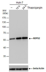
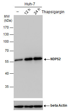
- NDP52 antibody detects NDP52 protein by western blot analysis. Un-treated (-) and treated (+, Thapsigargin treatment for 12hrs and 24hrs) Huh-7 whole cell extracts (30 £gg) were separated by 10% SDS-PAGE, and the membrane was blotted with NDP52 antibody (GTX630397) diluted by 1:500.The ACTB was used as internal control (GTX110564, 1:50000) shown at the bottom panel.
- Submitted by
- GeneTex (provider)
- Main image
- Experimental details
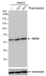
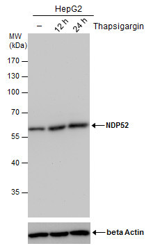
- NDP52 antibody detects NDP52 protein by western blot analysis. Un-treated (-) and treated (+, Thapsigargin treatment for 12hrs and 24hrs) HepG2 whole cell extracts (30 £gg) were separated by 10% SDS-PAGE, and the membrane was blotted with NDP52 antibody (GTX630397) diluted by 1:500.The ACTB was used as internal control (GTX110564, 1:50000) shown at the bottom panel.
- Submitted by
- GeneTex (provider)
- Main image
- Experimental details
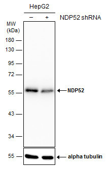
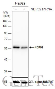
- Non-transfected (¡V) and transfected (+) HepG2 whole cell extracts (30 ?g) were separated by 10% SDS-PAGE, and the membrane was blotted with NDP52 antibody [GT1813] (GTX630397) diluted at 1:1000.
Supportive validation
- Submitted by
- GeneTex (provider)
- Main image

- Experimental details
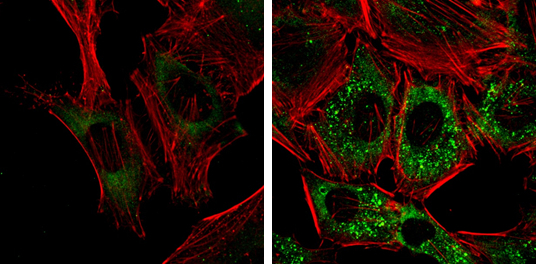
- NDP52 antibody [GT1813] detects NDP52 protein at autophagosome by immunofluorescent analysis. Samples: HeLa cells mock (left) and treated with 50£gM Chloroquine for 24 hr (right) were fixed in 4% paraformaldehyde at RT for 15 min.Green: NDP52 protein stained by NDP52 antibody [GT1813] (GTX630397) diluted at 1:1000.Red: Phalloidin, a F-actin marker.