 Explore
 Validate
 Learn
Learn Western blot
Antibody data
- Antibody Data
- Antigen structure
- References [0]
- Comments [0]
- Validations
- Western blot [1]
- Immunohistochemistry [1]
Submit
Validation data
Reference
Comment
Report error
- Product number
- TA319500 - Provider product page

- Provider
- OriGene
- Product name
- Rabbit polyclonal APC1 phospho S377 antibody (Phospho-specific)
- Antibody type
- Polyclonal
- Description
- Rabbit polyclonal APC1 phospho S377 antibody (Phospho-specific)
- Host
- Rabbit
- Conjugate
- Unconjugated
- Epitope
- ANAPC1
- Isotype
- IgG
- Antibody clone number
- NULL
- Vial size
- 100 µg
- Concentration
- 0.21 mg/mL
No comments: Submit comment
Supportive validation
- Submitted by
- OriGene (provider)
- Main image
- Experimental details
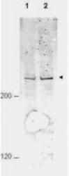
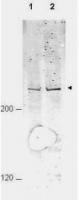
- WB using Anti-APC1 pS377 antibody shows detection of a band ~215 kDa corresponding to phosphorylated human APC1 (arrowhead). Lane 1 shows lysate from asynchronous cells. Lane 2 shows lysate from cells treated with nocodazole. While some phosphorylated APC1 is present in untreated cell, the amount of phosphorylated protein is increased in cell preparations arrested in mitosis. Primary antibody diluted to 1:1,000. Sea 1:10,000 dilution of IRDye800 conjugated Gt-a-Rabbit IgG [H&L].
- Validation comment
- WB
Supportive validation
- Submitted by
- OriGene (provider)
- Main image
- Experimental details
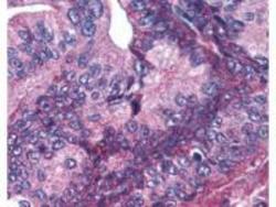
- Anti-APC1 pS377 antibody was used at 5.0 ug/ml to detect signal in a variety of tissues including multi-human, multi-brain and multi-cancer slides. This image shows moderate positive cytoplasmic and occasional nuclear staining of pancreatic carcinoma cells at 60X. Tissue was formalin-fixed and paraffin embedded. The image shows localization of the antibody as the precipitated red signal, with a hematoxylin purple nuclear counterstain.
- Validation comment
- IHC
