Explore
 Validate
 Learn
Learn Western blot
Antibody data
- Antibody Data
- Antigen structure
- References [0]
- Comments [0]
- Validations
- Western blot [2]
- Immunocytochemistry [1]
- Other assay [1]
Submit
Validation data
Reference
Comment
Report error
- Product number
- PA5-18434 - Provider product page

- Provider
- Invitrogen Antibodies
- Product name
- HEXIM1 Polyclonal Antibody
- Antibody type
- Polyclonal
- Antigen
- Synthetic peptide
- Description
- This antibody is predicted to react with mouse based on sequence homology.
- Reactivity
- Human, Mouse
- Host
- Goat
- Isotype
- IgG
- Vial size
- 100 µg
- Concentration
- 0.5 mg/mL
- Storage
- -20° C, Avoid Freeze/Thaw Cycles
No comments: Submit comment
Supportive validation
- Submitted by
- Invitrogen Antibodies (provider)
- Main image

- Experimental details
- PA5-18434 1) HeLa cell lysate and 2) recombinant HEXIM1 (3 ng). Detected by chemiluminescence.

- Submitted by
- Invitrogen Antibodies (provider)
- Main image

- Experimental details
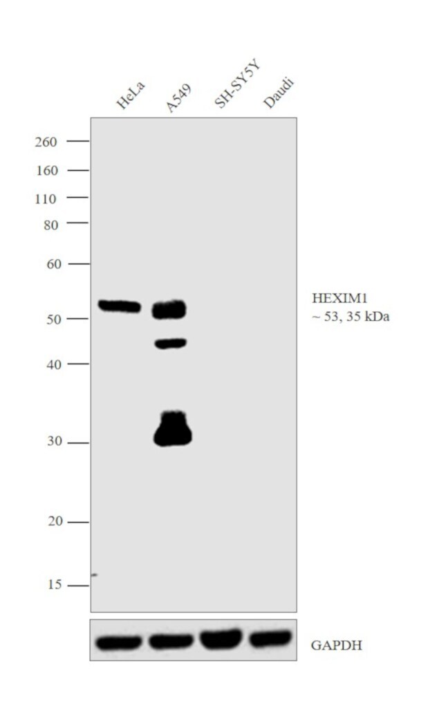
- Western blot was performed using Anti-HEXIM1 Polyclonal Antibody (Product # PA5-18434) and 53, 35 kDa bands corresponding to HEXIM1 was observed in HeLa, A549 and not in SH-SY5Y and Daudi which are reported negative for HEXIM1 expression. Modified whole cell extracts (1% SDS) (30 µg lysate) of HeLa (Lane 1), A549 (Lane 2), SH-SY5Y (Lane 3) and Daudi (Lane 4) were electrophoresed using Novex® NuPAGE® 4-12 % Bis-Tris gel (Product # NP0322BOX). Resolved proteins were then transferred onto a nitrocellulose membrane (Product # IB23001) by iBlot® 2 Dry Blotting System (Product # IB21001). The blot was probed with the primary antibody (0.3 µg/mL) and detected by chemiluminescence Rabbit anti-Goat IgG (H+L) Superclonal™ Secondary Antibody, HRP conjugate (Product # A27014, 1:4000 dilution) using the iBright FL 1000 (Product # A32752). Chemiluminescent detection was performed using Novex® ECL Chemiluminescent Substrate Reagent Kit (Product # WP20005).
Supportive validation
- Submitted by
- Invitrogen Antibodies (provider)
- Main image
- Experimental details
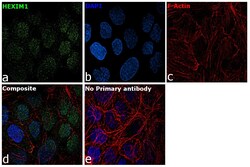
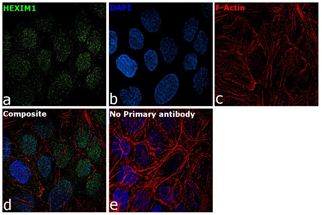
- Immunofluorescence analysis of HEXIM1 was performed using A-431 cells. The cells were fixed with 4% paraformaldehyde for 10 minutes, permeabilized with 0.1% Triton™ X-100 for 15 minutes, and blocked with 2% BSA for 1 hour at room temperature. The cells were labeled with HEXIM1 Polyclonal Antibody (Product # PA5-18434) at 5µg/mL in 0.1% BSA, incubated at 4 degree Celsius overnight and then labeled with Rabbit anti-Goat IgG (H+L) Secondary Antibody, Alexa Fluor® 488 conjugate (Product # A-11078) at a dilution of 1:2000 for 45 minutes at room temperature (Panel a: green). Nuclei (Panel b: blue) were stained with ProLong™ Diamond Antifade Mountant with DAPI (Product # P36962). F-actin (Panel c: red) was stained with Rhodamine Phalloidin (Product # R415, 1:300). Panel d represents the merged image showing nuclear localization. Panel e represents control cells with no primary antibody to assess background. The images were captured at 60X magnification.
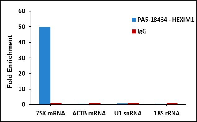
Supportive validation
- Submitted by
- Invitrogen Antibodies (provider)
- Main image

- Experimental details
- Detection of binding of endogenous HEXIM1 protein to specific RNA using Anti-HEXIM1 Antibody: RNA Immunoprecipitation (RIP) was performed using Anti-HEXIM1 Recombinant Rabbit Polyclonal Antibody (Product # PA5-18434, 5 µg) on whole cell lysate from 2 million HCT 116 cells. Normal Rabbit IgG was used as a negative IP control. RNA purified by RiboPure™ RNA Purification Kit (Product # AM1924) was analyzed by RT-PCR using the Power SYBR® Green RNA-to-CT™ 1-Step Kit (Product # 4389986) with RIP primer pairs over 7SK, ACTB mRNA, U1 snRNA and 18s rRNA. Data is presented as fold enrichment of the antibody signal versus the negative control IgG using the comparative CT method.